Download
1 / 1
30 likes | 167 Views
A 34-year old woman with acute pain in the left flank. K.R. van IJzendoorn, R.A.L Jacobs, K.L.J. Rademakers, L.M.C.L. Fossion. Department of Urology, Máxima medical center, Veldhoven, The Netherlands. Case report

E N D
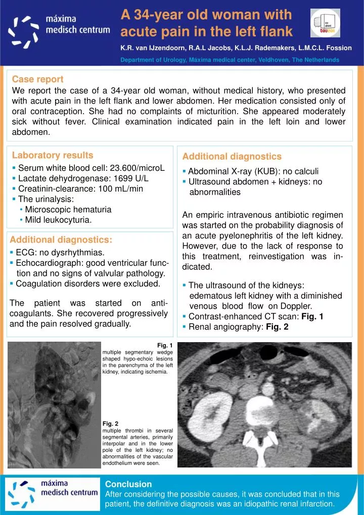
A 34-year old woman with acute pain in the left flank K.R. van IJzendoorn, R.A.L Jacobs, K.L.J. Rademakers, L.M.C.L. Fossion Department of Urology, Máxima medical center, Veldhoven, The Netherlands Case report We report the case of a 34-year old woman, without medical history, who presented with acute pain in the left flank and lower abdomen. Her medication consisted only of oral contraception. She had no complaints of micturition. She appeared moderately sick without fever. Clinical examination indicated pain in the left loin and lower abdomen. • Laboratory results • Serum white blood cell: 23.600/microL • Lactate dehydrogenase: 1699 U/L • Creatinin-clearance: 100 mL/min • The urinalysis: • Microscopic hematuria • Mild leukocyturia. • Additional diagnostics • Abdominal X-ray (KUB): no calculi • Ultrasound abdomen + kidneys: no abnormalities • An empiric intravenous antibiotic regimen was started on the probability diagnosis of an acute pyelonephritis of the left kidney. However, due to the lack of response to this treatment, reinvestigation was in-dicated. • The ultrasound of the kidneys: edematous left kidney with a diminished venous blood flow on Doppler. • Contrast-enhanced CT scan: Fig. 1 • Renal angiography: Fig. 2 • Additional diagnostics: • ECG: no dysrhythmias. • Echocardiograph: good ventricular func- tion and no signs of valvular pathology. • Coagulation disorders were excluded. The patient was started on anti-coagulants. She recovered progressively and the pain resolved gradually. Fig. 1 multiple segmentary wedge shaped hypo-echoic lesions in the parenchyma of the left kidney, indicating ischemia. Fig. 2 multiple thrombi in several segmental arteries, primarily interpolar and in the lower pole of the left kidney; no abnormalities of the vascular endothelium were seen. Conclusion After considering the possible causes, it was concluded that in this patient, the definitive diagnosis was an idiopathic renal infarction.