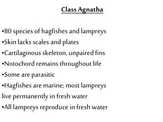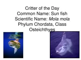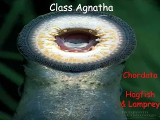
Class Agnatha
Lepidosuauromorpha. Class Osteichthyes. Class Chondrichthyes. Class Mammalia. Testudines. Crocodilia. Dinosauria. Class Amphibia. Class Agnatha. Class Aves. Archosauromorpha. Diapsida. Eureptilia. Sauropsid opening. Amniota. Amniote egg present. Tetrapoda. Girdles and limbs.

Class Agnatha
E N D
Presentation Transcript
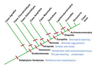
Lepidosuauromorpha Class Osteichthyes Class Chondrichthyes Class Mammalia Testudines Crocodilia Dinosauria Class Amphibia Class Agnatha Class Aves Archosauromorpha Diapsida Eureptilia Sauropsid opening Amniota Amniote egg present Tetrapoda Girdles and limbs Teleostomi Vertebrates with endochondral bone Gnathostomata The jaw-bearing vertebrates Subphylum Vertebrata Vertebrae and cephalization
Bone formation Can occur two ways. 1) Endochondral Bone Also called cartilage replacement bone. During ossification (=mineralization with CaPO4) a cartilage model is replaced 2) Dermal Bone Think of this as skin bone Dermal bone Forms in the dermis of the skin No cartilage model or precursor
Extinct agnathans Several groups were armored with dermal bone. Internal skeleton is cartilage. Dermal bone is phylogentically older. Teleostomi Bony fish plus tetrapods Internal skeleton is endochondral bone. Sheets of dermal bone incorporated into skull roof. Dermal bone used as armor in many forms.
Lepidosuauromorpha Class Osteichthyes Class Chondrichthyes Class Mammalia Testudines Crocodilia Dinosauria Class Amphibia Class Agnatha Class Aves Archosauromorpha Diapsida Eureptilia Sauropsid opening Amniota Amniote egg present Tetrapoda Girdles and limbs Teleostomi Vertebrates with endochondral bone Gnathostomata The jaw-bearing vertebrates Subphylum Vertebrata Vertebrae and cephalization dermal bone
Characteristics of Tetrapoda Girdles and limbs A very primitive amphibian Pectoral girdle Pelvic girdle Fore limb Hind limb Limbs of all tetrapods have the same basic components.
Lepidosuauromorpha Class Osteichthyes Class Chondrichthyes Class Mammalia Testudines Crocodilia Dinosauria Class Amphibia Class Agnatha Class Aves Archosauromorpha Diapsida Eureptilia Sauropsid opening Amniota Amniote egg present Tetrapoda Girdles and limbs Teleostomi Vertebrates with endochondral bone Gnathostomata The jaw-bearing vertebrates Subphylum Vertebrata Vertebrae and cephalization
Always has three embryonic membranes (1) Amnion (2) (3)
Synapsids Anapsids Lepidosuauromorpha Class Osteichthyes Class Chondrichthyes Class Mammalia Testudines Crocodilia Dinosauria Class Amphibia Class Agnatha Class Aves Archosauromorpha Diapsida Eureptilia Sauropsid opening Amniota Amniote egg present Tetrapoda Girdles and limbs Teleostomi Vertebrates with endochondral bone Gnathostomata The jaw-bearing vertebrates Subphylum Vertebrata Vertebrae and cephalization
po sq j q po = postorbital sq = squamosal j = jugal po sq j q = quadrate q (and mammals) po sq j q
Skeletal system Major components Skull, vertebrae, ribs Axial skeleton - Appendicular skeleton - girdles limbs Function Lever arms in locomotion, Protection of internal structures A mineral “bank” Produce/support structures used in communication.
Skeletal system Cranial skeleton Postcranial skeleton Major components Skull, vertebrae, ribs Axial skeleton - girdles, limbs, Appendicular skeleton - The vertebrate skull is produced by three major components: The splanchnocranium Forms the jaws, gill arches, hyoid, Cartilages at the anterior end of the trachea, glottis, epiglottis and larynx The chondrocranium Forms the braincase The dermatocranium Forms the skull roof, armor on top of skull
Cranial skeleton Skeletal system Major components Skull, vertebrae, ribs Axial skeleton - The vertebrate skull is produced by three major components: The chondrocranium Cartilaginous in Agnathans and Chondrichthyes This structure is replaced by endochondral bone in all Teleostomi
The chondrocranium is hidden by dermatocranium
The chondrocranium Is still visible in the cat As the skull bones on which the brain rests: Basisphenoid (2) Presphenoid (22) Alisphenoid (no #) And the bones surrounding the foramen magnum: The occipitals (16)
Skeletal system Major components of the skull 1) The chondrocranium the brain basket 2) The splanchnocranium Forms the jaws, gill arches, hyoid,
chondrocranium Splanchnocranium is in blue Chondrocranium is in black
Branchial Arches Jaws: Hyoid Arch: 17 - palatoquadrate cartilage 9 - hyomandibular 6 - epibranchial 5 - ceratohyal 4 - cerabranchial 11 - Meckle’s cartilage Serial homology is an important concept
The splanchnocranium Is represented in the cat skull by dermal bone That grows around the embyrological palatoqudrate and mandibular cartilages: Maxilla premaxilla palatine pterygoid Dentary
Skeletal system Major components of the skull 1) The chondrocranium Brain basket 2) The splanchnocranium Jaws and gill arches 3) The dermatoocranium Forms the skull roof, armor on top of skull The shark has no dermatocranium
Dermal bones of the skull roof are the most obvious elements in dorsal view nasal frontal parietal, squamosal, jugal,
It is the dermatocranium that is fenestrated within the amniota.
embryological source of skull elements in the mammal
Skull: Anterior View E.N. Marieb 6th ed. Figure 7.2a Figure 7.2a
Lateral view of human skull E.N. Marieb 6th ed. Figure 7.3a E.N. Marieb 6th ed. Figure 7.3a
E.N. Marieb 6th ed. Figure 7.4a Figure 7.4b
Inferior Veiw of the Human Skull E.N. Marieb 6th ed. Figure 7.4a Figure 7.4a
Mandible and Its Markings E.N. Marieb 6th ed. Figure 7.8a Figure 7.8a
Developmental Aspects: Fetal Skull • Skull bones such as the mandible and maxilla are unfused E.N. Marieb 6th ed. Figure 7.33 Figure 7.33
Cleft Palate Lindsay Biggs http://www.biomedcentral.com/1471-2350/5/15/figure/f1
Causes: Gene mutations Environmental factors Maternal diet Medication and drug uses Ethnic background • Background: • Palate – roof of the mouth • functions as a barrier between the nasal and oral portions of the respiratory tract • Cleft – fissure or opening • Incorrect joining of the mouth tissues • Most common congenital deformity of the head and face • Related Issues: • Feeding • Speech • Hearing • Facial growth http://www.leap-foundation.org/cleft-lip-and-palate-information/
Cleft Palate Types: Incomplete – a gap that does not run all the way from the lip and mouth and into the nose Complete – a gap that goes all the way from the lip and mouth into the nose Unilateral – occurs on only one side of the upper lip Bilateral – occurs on both sides of the upper lip Microform – a form of incomplete cleft that is very minor, such as a groove or notch http://bestpractice.bmj.com/best-practice/monograph/675/resources/image/bp/5.html
Embryology The face undergoes major transformations between the 5th and 10th week of gestation Stomodeum – precursor of the mouth Frontalnasal prominence – a midline swelling that forms the bridge of the nose Nasal pits – invaginations that become the nostrils • Mandibular prominences – form the lower lip and madible https://missinglink.ucsf.edu/restricted/lm/CongenitalAnomalies/CleftLipPalate.html
Treatments Options First 6 months - initial repair of the lip and nose 12-18 months – palate repair 7-9 years – repair of the cleft in the gum line Additional surgeries may be necessary to improve speech and repair improper jaw growth