Pneumonia
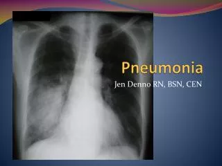
Pneumonia. Jen Denno RN, BSN, CEN. Pneumonia . Vaccination Percent of adults 65 years and over who had ever received a pneumococcal vaccination: 59% Health Care Use Hospital inpatient care Number of discharges: 1.1 million Average length of stay: 5.2 days Nursing home care

Pneumonia
E N D
Presentation Transcript
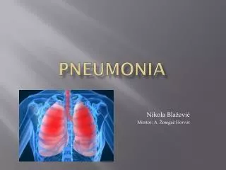
Pneumonia Jen Denno RN, BSN, CEN
Pneumonia • Vaccination • Percent of adults 65 years and over who had ever received a pneumococcal vaccination: 59% • Health Care Use • Hospital inpatient care • Number of discharges: 1.1 million • Average length of stay: 5.2 days • Nursing home care • Number of residents with pneumonia: 33,700 • Percent of residents with pneumonia: 2.3% • Mortality • Number of deaths: 50,774 • Deaths per 100,000 population: 16.5
VIRAL • Viral infections are characterized by the accumulation of mono- nuclear cells in the submucosa • and perivascular space, resulting in partial obstruction of the airway. Patients with these infections present with wheezing and crackles. • Disease progresses when the alveolar type II cells lose their structural integrity and surfactant production is diminished, a hyaline membrane forms, and pulmonary edema develops.
Bacterial • In bacterial infections, the alveoli fill with proteinaceous fluid, which triggers a brisk influx of red blood cells (RBCs) and polymorphonuclear (PMN) cells (red hepatization) followed by the deposition of fibrin and the degradation of inflammatory cells (gray hepatization). During resolution, intra-alveolar debris is ingested and removed by the alveolar macrophages. This consolidation leads to decreased air entry and dullness to percussion; inflammation in the small airways leads to crackles.
Four stages of lobar pneumonia have been described. • In the first stage, which occurs within 24 hours of infection, the lung is characterized microscopically by vascular congestion and alveolar edema. Many bacteria and few neutrophils are present. • The stage of red hepatization (2-3 d), so called because of its similarity to the consistency of liver, is characterized by the presence of many erythrocytes, neutrophils, desquamated epithelial cells, and fibrin within the alveoli. • In the stage of gray hepatization (2-3 d), the lung is gray-brown to yellow because of fibrinopurulentexudate, disintegration of RBCs, and hemosiderin. • The final stage of resolution is characterized by resorption and restoration of the pulmonary architecture. Fibrinous inflammation may lead to resolution or to organization and pleural adhesions.
In interstitial pneumonia, patchy or diffuse inflammation involving the interstitium is characterized by infiltration of lymphocytes and macrophages. The alveoli do not contain a significant exudate, but protein-rich hyaline membranes similar to those found in adult respiratory distress syndrome (ARDS) may line the alveolar spaces. • Bacterial superinfection of viral pneumonia can also produce a mixed pattern of interstitial and alveolar airspace inflammation.
Klebsiella • How do you get it? • What does it look like? • What happens as the condition • spreads?
They gain entry into the body by eating unwashed vegetables and drinking contaminated water. • Most of the time, a Klebsiellapneumoniae infection is very common in patients with underlying diseases like diabetes, chronic lung diseases, chronic alcoholics, etc. It is mostly a nosocomial infection that occurs in hospitalized patients with weakened immune system. • Once Klebsiellapneumoniae enters the lungs, it causes many destructive changes in the lungs. It leads to necrosis, inflammation, hemorrhage, etc. of the lung tissues. This leads to production of a very thick, jelly like mucus that is called 'currant jelly sputum'. The rapid destruction of the lung tissues is the distinguishing factor for Klebsiellapneumoniae infection. Initially, Klebsiellapneumoniae will cause a sudden high fever. This fever is generally more than 103ºF. The fever is accompanied by other symptoms like chills and dizziness. The patient will also cough up the thick currant jelly sputum. This sputum may show streaks of blood. • As the condition spreads, it leads to formation of abscess. These abscesses are dead tissue pockets that contain millions of Klebsiellapneumoniae bacteria. Formation of abscesses cause the lungs to stick with the connective tissues surrounding them. This may lead to collapsed lungs in some patients. Soon, the infection spreads to the upper respiratory tract. When the infection spreads, it causes severe airway congestion. This leads to a foul-smelling nasal discharge.