OSTEOPOROSIS
OSTEOPOROSIS. Dr. Edward Warren Chair, Geriatrics Carolinas Campus May 2012. Objectives. Define osteoporosis. Apply knowledge of risk factors of osteoporosis to patient evaluation and management. Discuss diagnosis of osteoporosis. Discuss fractures associated with osteoporosis.

OSTEOPOROSIS
E N D
Presentation Transcript
OSTEOPOROSIS Dr. Edward Warren Chair, Geriatrics Carolinas Campus May 2012
Objectives • Define osteoporosis. • Apply knowledge of risk factorsof osteoporosis to patient evaluation and management. • Discuss diagnosis of osteoporosis. • Discuss fractures associated with osteoporosis. • Treat osteoporosis optimally. • Prevent osteoporosis effectively.
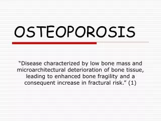
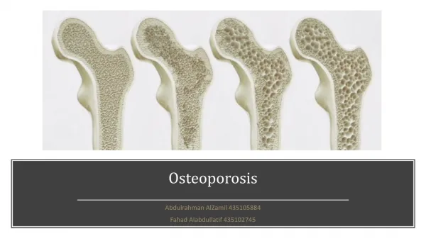
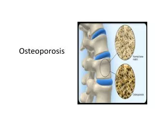
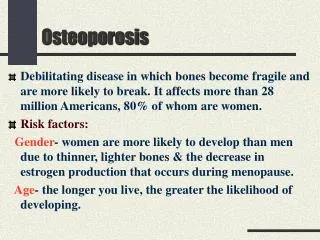
Osteoporosis • A reduction in bone mass with an increased risk and ease of fractures • Loss of bone tissue results in a deterioration of skeletal microarchitecture
Osteoporosis • Increased “porosity” of bone • Prevalence = 10,000,000 in the US only a minority of these are diagnosed and treated
Osteopenia • Osteopenia=low bone density • “penia” from Greek “penes”, related to “penury” meaning “poor”. • Osteopenic women have more than 50% of the fractures postmenopause. • Prevalence = 18,000,000 in the US
PrevalenceUnderdiagnosed & Undertreated in ALL! • Osteopenia in Caucasians & Asians • Women 52% • Men 35% • Osteoporosis in Caucasians & Asians • Women 20% • Men 7% • Hispanic women are catching up fast! • Blacks have 40% less fractures, but more fatalities!
Bone Remodeling • Bone repairs itself by constant, active remodeling • bone reabsorption (osteoclasts) • bone formation (osteoblasts) • The remodeling cycle may become unbalanced • after menopause • with aging in both men and women • bone reabsorption rate exceeds bone formation rate: weeks vs months • net bone loss is predominantly cancellous (trabecular) bone.
Risk Factors • Personal fracture • 1° relative fracture • Female gender • Advanced age • Caucasian/Asian • Low Ca intake • Vitamin D deficiency • Low hormone levels • Tobacco abuse • Alcohol Abuse • Sedentary Lifestyle • Frailty • Ectomorphic • Certain medications • Certain diseases
Risk Factors: Medications • Anticonvulsants • Warfarin • Glucocorticoids • Lithium • Neuroleptics • Medroxyprogesterone • Excess levothyroxine • Excess Vitamin A • Excess Vitamin D • PO4 binding antacids: Aluminum hydroxide • Aromatase Inhibitors & GnRH Antagonists (for CA prostate) • Immunosuppressants& Cytotoxins (i.e. MTX)
Risk Factors: Diseases • Cushing’s Disease • Addison’s Disease • 1° Hyperparathyroidism • Hyperthyroidism • Hypogonadism • Multiple myeloma • Leukemias • Sickle Cell • Osteomalacia • Paget’s Disease • COPD • Vitamin D deficiency • Malabsorption syndromes • Malnutrition • RA, Lupus • Diabetes Mellitus • Renal Failure
Risk Factors: Ca & Vitamin D • Calcium, vitamin D & parathyroid hormone maintain bone homeostasis. • Low Ca in diet or malabsorption of it leads to 2° hyperparathyroidism. • PTH is excreted when serum Ca is low, leading to increased bone resorption, decreased renal Ca excretion, and increased renal production of 1,25 dihydroxy-vitamin D. • Vitamin D increases absorption of Ca and P from the gut, and inhibits PTH synthesis.
Clinical Manifestations • Hip fractures • Vertebral and other Osteoporotic Fractures • Wrist fractures • Most any fracture you can think of • pelvis, head of the humerus, etc. • 1,500,000 fractures yearly in the US
HIP Fractures • Incidence doubles q5 years after age 70. • 300,000 hip fractures yearly in the US • Likelihood for a 50 year old Caucasian to fracture a hip is: 14% women, 5% men • African Americans are half as likely. • Only ⅓ have a full recovery. • 20% live in a nursing home lifelong.
HIP Fractures Complications: • Deep venous thrombosis • Pulmonary embolism • 5 – 20% mortality rate • 66% suffer permanent mobility loss • Doubles risk of future fractures.
Vertebral Compression Fractures • 700,000 yearly in the US • Many are asymptomatic • Height loss • Kyphosis • Back pain
Vertebral Compression Fractures • Thoracic compression fractures • Restrictive lung disease • Lumbar compression fractures • abdominal distention • early satiety • constipation
PEARL THE MOST IMPORTANT PART OF TREATMENT IS DIAGNOSIS.
Evaluation • Comprehensive H&PE as ALWAYS! • Measure bone mineral density: BMD • Assess for secondary causes of bone loss: UGH!
H&PE • FH: fractures, especially at early age • SH: tobacco, ethanol, activity, diet • PMH: prior fractures, risk factors, fragility fractures (with minimal force), vitamin D & calcium use • Elderly fall risk: balance, orthostatic hypotension, debility, poor vision, poor hearing, poor cognition
H&PE • BMI < 20 • kyphoscoliosis • vertebral point tenderness • height loss
Bone Mineral Density • Dual-energy X-ray Absorptiometry (DEXA, DXA) • highly accurate, standard in most centers • Single-energy X-ray Absorptiometry (SEXA, SXA, SEX?) • less accurate, less available • Quantitative CT • expensive, more radiation, less reproducible • gives true density • Ultrasound • cheap and mobile
Bone Mineral Density: DXA • Preferred method of measurement • Can measure hip, anterior-posterior spine, lateral spine, and wrist • Cost = $200 to $300 • Covered by Medicare and Medicaid at 24 month intervals on anyone
Bone Mineral Density: DXA • Two dimensional technique. • Two x-ray energies are used to measure the mineralization in an area. The mineral content is divided by this area. • Small people are rated spuriously low. • Machines vary from each other = problem
Bone Mineral Density: DXA • STASTICS TO THE RESCUE • A Bell-shaped curve is formed for each population by age, gender, and race. • T-score compares one to 30 year olds matched for gender and race. • Z-score compares one to age, gender, and race matched controls.
T-score graph The WHO has defined the following categories based on bone density in white women: Normal bone: T-score > -1 Osteopenia: -1 > T-score > -2.5 Osteoporosis: T-score < -2.5
National Osteoporosis Foundation Recommendations for DXA • Women > age 65 & Men > age 70 • Estrogen-deficient women with clinical risks • Osteopenia or vertebral fracture on x-ray • Fragility fractures • Patients with illness or medication that imparts risk > age 50 • Glucocorticoid treatment ≥7.5 mg of prednisone duration of therapy > 3 months • Primary hyperparathyroidism • Monitoring response to an FDA-approved medication for osteoporosis
Monitoring with DXA • Repeat BMD evaluations at > 2 year intervals since years are needed for any changes • Significant changes are 4% in spine and 6% in hips • Treatments do not often produce changes large enough to detect reliably • In those below age 50, use Z-scores. If < -2.0, then they are “below expected range”, and would need additional clinical criteria for a diagnosis.
FRAX • This is the WHO fracture risk algorithm • www.shef.uk/FRAX/ • Enter clinical data and get a fracture risk. • The National Osteoporosis Foundation advises treatment for 3% risk of hip fracture or 20% risk of osteoporotic fractures.
Screening for Secondary Causes • ↑ Ca hyperparathyroidism or malignancy • ↑ iPTH hyperparathyroidism • ↓ iPTH malignancy (↑ PTHrP suggests humoral hypercalemia of malignancy) • ↓ Ca osteomalacia, malnutrition, or malabsorption
Screening for Secondary Causes • Cushing’s suspected dexamethasone suppression test • Malnutrition suspected albumen, prealbumen, cholesterol, CBC, vitamin B12, folic acid, Fe, TIBC if low, then further evaluation needed • Myeloma suspected serum protein electrophoresis, urine electrophoresis for light chains, and bone marrow exam • X-rays are for fracture documentation only.
Preventing and Treating Osteoporosis • Weight-bearing exercise: lifelong • Smoking cessation: lifelong • Ethanol moderation: lifelong • Calcium and vitamin D: dietary, sunlight, lifelong • Estrogen replacement • Bisphosphonates • Selective estrogen receptor modulators • Calcitonin • Others
Treatment Criteria • T-score < -2.5 • Any postmenopausal woman with risk factors regardless of T-score
Risk Factor Reduction • Glucocorticoids can be titrated to minimal effective doses. • Thyroid hormone doses need to be as low as needed to normalize TSH. • Smoking cessation. • Ethanol moderation.
Fall Risk Factor Reduction • Ethanol avoidance. • Reduce or eliminate sedating medications. • Protect against orthostatic hypotension. • Address nocturia. • Fix environmental hazards: wires, rugs, furniture, socks • Vision correction
Exercise • Decrease in physical activity or immobilization decline in bone mass • Walking, a weight-bearing exercise, can be recommended for all adults • Exercise 2% bone mass increase at best. NNT = 1600 • Start slowly and gradually increase the number of days and time spent walking each day (at least 3 days weekly) • Improves coordination, balance, and strength.
DOE’s vs POE’s • DOE = disease oriented evidence • 2% increase in bone mineral density • POE = patient oriented evidence • how many fractures are prevented
NNT • Alendronate reduces vertebral compression fractures 44%: relative risk. • Shun, reject, ignore relative risk. • Liars figure and figures lie. • At worst relative risk is used to fool you into believing something is better than it really is. • At best it is being presented by someone who does not know any better.
NNT • Alendronate reduces vertebral compression fractures 44% in 3 years • Placebo 3.9% fractures: alendronate 2.2% • Number Needed to Treat • 1.7% absolute reduction • 1.7/100 patients reciprocal • NNT of 59 patients to prevent 1 fracture/3 years
NNT • “NNT” is well ensconced in the medical parlance and can be searched for on the internet with a drug name to find the value easily. • Bisphosphonates reduce hip fractures about 40%, relative risk. • About 1% of people over 65 have hip fractures yearly • Absolute reduction is thus 0.4% or 4/1000 • The reciprocal of 0.004 is NNT = 250 • Is it worth $250,000 to prevent one hip fracture a year?
Calcium & Vitamin D • Calcium and vitamin D maintain or increase bone density in postmenopausal women & help prevent hip and nonvertebral fractures in all older adults. • Calcium has no benefit on fracture rate used alone. • Vitamin D: NNT = 45 to prevent a hip fracture over 3 years. • 1200 mg / day of calcium: men 65 years and older & postmenopausal women. • 1000 IU / day of vitamin D3: regardless of sunlight exposure to offset skin changes that efficient use of UV light to synthesize vitamin D precursors. (I start with 4000 units qd.) • Calcium has no harmful cardiac effects.
Estrogen Replacement • Estrogen produces bone loss protection. • All benefit is lost in 10 years if stopped. • 10,000 treated women in 1 year • 8 extra MI’s 18 extra DVT’s • 8 extra CVA’s 6 fewer CA colon • 5 fewer hip fractures 44 fewer fractures • neutral death rate (voided by progesterone) • NNT 385 for hip fractures over 3 years
Estrogen Replacement • SERMS (selective estrogen response modulators) • Tamoxifen reduces bone turnover and CA breast, but it increases uterine CA. • Raloxifene reduces vertebral collapse as well as CA breast. NNT is 22 in 4 years for a vertebral fracture.
Bisphosphonates • Structurally related to pyrophosphates which are incorporated into bone matrices where resorption is active. • Impair osteoclast function. • Poorly absorbed: bioavailability only 5%. • Reduce osteoclast number by apoptosis • NNT for hip fracture 77 – 91 in 3 years • NNT for vertebral fracture 13 – 20 in 3 years • Side effects: dyspepsia & esophageal perforation • Incapacitating bone pain, arthralgia, and myalgia can occur any time.
Bisphosphonates: Alendronate • In comparison with placebo: • bone density of spine (8%) & hip (3.5%) – DOE’s • vertebral & hip fracture rate by 50% at 2 years • NNT 37 for vertebral fractures in 3 years – POE • Dosing for Prevention: Once-weekly 70 mg po: $102/month • Side effects • GI: abdominal pain, dyspepsia, esophagitis, nausea, vomiting, diarrhea • Musculoskeletal pain
Bisphosphonates: Zoledronic Acid • Most potent. Given by yearly IV infusion. • In comparison with placebo: • bone density of spine (5.1%) & hip (3.5%) – DOE’s • vertebral & hip fracture rate by 70% and 41% respectively at 3 years • NNT 107 for vertebral fractures in 3 years – POE • NNH (number needed to harm) = 100 • Cost $1250/year • Side effects • GI: abdominal pain, dyspepsia, esophagitis, nausea, vomiting, diarrhea • Musculoskeletal pain
Other Bisphosphonates • Risedronate • Approved for osteoporosis: 35 mg po weekly: $130/month • In comparison with placebo: • bone density of spine (5.4%) & hip (1.6%) – DOE’s • new vertebral & hip fracture rate of 50% – relative risk • GI side effects • Ibandronate: similar: $470/3 months • Benefits lost 1 year after stopping • NNT = 13 – 20 over 3 years for vertebral fracture • Death rates equal in drug and placebo groups