Download
1 / 18
190 likes | 391 Views
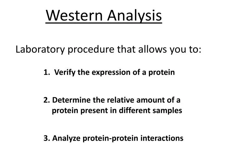
Western Analysis. Laboratory procedure that allows you to: . 1. Verify the expression of a protein. 2. Determine the relative amount of a protein present in different samples. 3. Analyze protein-protein interactions. Two Main Types of Westerns. 1. Denaturing (Most Commonly Used).

E N D
Western Analysis Laboratory procedure that allows you to: 1. Verify the expression of a protein 2. Determine the relative amount of a protein present in different samples 3. Analyze protein-protein interactions
Two Main Types of Westerns 1. Denaturing (Most Commonly Used) - SDS-PAGE 2. Non-Denaturing - Native PAGE
SDS-PAGE Western Blot Method Cell Lysis by Detergents and Sonication Cell Removal Cells in Culture Human Cells Containing Protein SDS or LDS - - - - - - - - - - Heat Denaturation of Proteins - - - - - Detergents Bind Proteins Load Proteins on Gel - - - - Apply Electric Current - - + Proteins Separate by Size
Transfer or Blot Protein from Gel to Nitrocellulose and/or PVDF Membrane Block Membrane with Non-Specific Proteins Incubate Membrane with 1o Antibody - - - - - - - - - - - - 1o Antibody is a Rabbit Anti-Human b-Actin Antibody - - - - - - - - 1o Antibody Binds Antigen (i.e. Protein of Interest) Non-Specific Proteins Bind to Unbound Regions of Membrane
Add HRP-Conjugated 2o Antibody 2o Antibody is a Goat Anti-Rabbit-HRP-Conjugated Antibody Add Chemiluminescent Substrate Luminol Light Detected by Film HRP - - - - - - - - - - - -
Magic Mark XP Western Protein Standard kDa 60 50 45 b-Actin 40 30 20 Invitrogen.com
We will be using 4-12% BisTris Gels today because our protein of interest is 45 kDa in size. For larger proteins (>100 kDa) this type of gel would not be appropriate because the resolution of large proteins on a 4-12% BisTris gel is poor.
Magic Mark XP Western Protein Standard kDa 60 50 45 b-Actin 40 30 20 Invitrogen.com QUESTION If we wanted to detect another human protein that was 60 kDa on this blot at the same time as beta actin could we? Why would it be wise to use a primary antibody against this protein that was generated in a rabbit?
Some Drawbacks of Western Blotting 1. Many steps where errors can occur 2. Large amount of sample needed (5-50 mg) 3. Accurate quantitation is very difficult 4. Time consuming protocol
Western Blot Protocol Sample Preparation A) Add 10 mg of protein to 5 ml of 4X LDS Loading Buffer plus 2.5 ml of 10X Reducing Agent. Then add purified water to a total volume of 25 ml. For example: If your total protein concentration is 2.0 mg/ml, you would need 5 ml of total protein to equal 10 mg. So you would mix: 5 ml of protein 5 ml of 4X LDS Loading Buffer 2.5 ml 10X Reducing Agent 12.5 ml purified water. B) Heat sample mixture at 70oC for 10 minutes.
2. Electrophoresis A) While protein samples are heating, assemble electrophoresis unit. Demonstration XCellSureLock Mini-Cell, Invitrogen • Load Gel • -Molecular weight marker and protein samples Invitrogen.com Demonstration Invitrogen.com
C) Add 500 ml Antioxidant to top chamber to maintain proteins in a reduced state and ensure optimal band sharpness. D) Run gel at 180V for 45 minutes
3. Transfer A) Soak marked (for orientation) nitrocellulose (or PVDF) membrane in transfer buffer containing 10% Methanol at least 10 minutes prior to transfer. When gel run is complete, turn off power source, remove gel from pre-cast plates, place transfer buffer-soaked filter paper sheet on top of gel, remove gel from plate, and place on top of membrane blotting pads that have been removed of bubbles. C) Place membrane on top of gel and cover with another transfer buffer-soaked filter paper sheet and blotting pads to fill the transfer chamber. Demonstration Blotting Pad Invitrogen.com D) Add 500 ml Antioxidant and run transfer at 30V for 1 hour.
4. Blocking A) Remove membrane from transfer chamber and incubate in 5% Blotto [5% powdered milk in TBS-Tw (1X TBS, 0.1% Tween 20)] at room temperature for 30 minutes with slow shaking. 5. Primary Antibody Incubation A) Prepare a 1:1000 dilution of primary antibody (Rabbit Anti-Human b-Actin) in 5% Blotto. B) Incubate membrane in primary antibody solution overnight at 4oC with gentle rocking.
6. Membrane Washing A) Wash membrane 3 x 5 minutes each in TBS-Twwith gentle shaking at room temperature. 7. Secondary Antibody Incubation A) Prepare a 1:5000 dilution of secondary antibody (Goat Anti-Rabbit IgG-HRP) in 5% Blotto. B) Incubate membrane in secondary antibody solution for 30 minutes at room temperature with gentle shaking. QUESTION Why are we using Goat Anti-Rabbit IgG-HRP as our secondary antibody? 8. Repeat Membrane Washing – See step 6
9. Visualization of Protein of Interest A) Place membrane protein side up on saran wrap on a flat surface. B) Quickly add 50 ml of ECL solution B to 2 ml of ECL solution A, mix, and add directly to membrane. C) Incubate in the dark for 3-5 minutes, remove excess solution, and place membrane protein side down onto a new piece of saran wrap. D) Close saran wrap around membrane, tape to film cassette and expose film in the dark room for 30 seconds to 1 minute. E) Develop film & identify protein of interest.
Magic Mark XP Western Protein Standard kDa 60 50 45 b-Actin 40 30 20