Download
1 / 70
700 likes | 721 Views
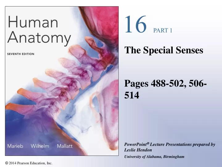
16 PART 1. The Special Senses Pages 488-502, 506-514. The Special Senses. Taste, smell, sight, hearing, and balance Touch —a large group of general senses Special sensory receptors Localized —confined to the head region Receptors are not free endings of sensory neurons

E N D
16 PART 1 The Special Senses Pages 488-502, 506-514
The Special Senses • Taste, smell, sight, hearing, and balance • Touch—a large group of general senses • Special sensory receptors • Localized—confined to the head region • Receptors are not free endings of sensory neurons • Special receptor cells • Are neuronlike epithelial cells or small peripheral neurons • Transfer sensory information to other neurons in afferent pathways
The Chemical Senses: Taste and Smell • Taste—gustation • Smell—olfaction • Receptors—classified as chemoreceptors • Respond to chemicals • Food dissolved in saliva • Airborne chemicals that dissolve in fluids of the nasal mucosa
Taste—Gustation • Taste receptors • Occur in taste buds • Most are found on the surface of the tongue • Located within tongue papillae • Two types of papillae (with taste buds) • Fungiform papillae • Vallate papillae
Taste Buds • Collection of 50–100 epithelial cells • Contain two major cell types • Gustatory epithelial cells supporting cells • Basal epithelial cells gustatory cells • Contain long microvilli—extend through a taste pore to the surface of the epithelium • Cells in tastebuds replaced every 7–10 days
Connective tissue Epiglottis Gustatory hair Taste fibers of cranial nerve Palatine tonsil Lingual tonsil Vallate papilla Stratified squamous epithelium of tongue Basal epithelial cells Gustatory epithelial cells Taste pore Fungiform papillae Taste bud (a) Taste buds associated with fungiform and vallate papillae (b) Enlarged section of a vallate papilla (c) Enlarged view of a taste bud (micrograph, 160X) Taste Buds Figure 16.1
Smell (Olfaction) • Olfactory receptors are part of the olfactory epithelium • Olfactory epithelium is pseudostratified columnar and contains three main cell types • Olfactory sensory neurons • Supporting epithelial cells • Basal epithelial cells
Smell (Olfaction) • Cell bodies of olfactory sensory neurons • Located in olfactory epithelium • Have an apical dendrite that projects to the epithelial surface • Ends in a knob from which olfactory cilia radiate • Olfactory cilia act as receptive structures for smell • Mucus captures and dissolves odor molecules
Smell (Olfaction) • Axons of olfactory epithelium • Gather into bundles—filaments of the olfactory nerve • Pass through the cribriform plate of the ethmoid bone • Attach to the olfactory bulbs and synapse with mitral cells • Mitral cells transmit impulses along the olfactory tract to • Limbic system • Piriform lobe of the cerebral cortex
Olfactory epithelium Mitral cell (output cell) Olfactory tract Glomeruli Olfactory bulb Olfactory tract Cribriform plate of ethmoid bone Olfactory bulb Filaments of olfactory nerve Lamina propria connective tissue Nasal conchae Olfactory gland Axon Basal cell Olfactory sensory neuron Route of inhaled air Olfactory epithelium Supporting cell (a) Dendrite Olfactory cilia Mucus Route of inhaled air containing odor molecules (b) Olfactory Receptors Figure 16.3
The Eye and Vision • Visual organ—the eye • 70% of all sensory receptors are in the eyes • 40% of the cerebral cortex is involved in processing visual information • Anterior one-sixth of the eye’s surface is visible
Accessory Structures of the Eye • Eyebrows—coarse hairs on the superciliary arches • Eyelids (palpebrae)—separated by the palpebral fissure • Meet at the medial and lateral angles (canthi) • Lacrimal caruncle—reddish elevation at the medial canthus • Tarsal plates—connective tissue within the eyelids • Tarsal glands—modified sebaceous glands
Levator palpebrae superioris muscle Orbicularis oculi muscle Eyebrow Tarsal plate Palpebral conjunctiva Tarsal glands Cornea Palpebral fissure Eyelashes Bulbar conjunctiva Conjunctival sac Orbicularis oculi muscle (b) Lateral view; some structures shown in sagittal section Accessory Structures of the Eye • Conjunctiva—transparent mucous membrane • Palpebral conjunctiva • Bulbar conjunctiva • Conjunctival sac Figure 16.4b
Lacrimal sac Lacrimal gland Excretory ducts of lacrimal glands Lacrimal punctum Lacrimal canaliculus Nasolacrimal duct Inferior meatus of nasal cavity Nostril Accessory Structures of the Eye • Lacrimal apparatus—keeps the surface of the eye moist • Lacrimal gland—produces lacrimal fluid • Lacrimal sac—fluid empties into nasal cavity Figure 16.5
Extrinsic Eye Muscles • Six muscles that control movement of the eye • Originate in the walls of the orbit • Insert on outer surface of the eyeball • Annular ring—origin of the four rectus muscles • The six extrinsic eye muscles are • Lateral rectus and medial rectus • Superior rectus and inferior rectus • Superior oblique and inferior oblique
Superior oblique muscle Trochlea Trochlea Superior oblique Superior rectus Superior oblique tendon Superior rectus muscle Medial rectus Lateral rectus Lateral rectus muscle Inferior rectus Inferior oblique Common tendinous ring Inferior rectus muscle Inferior oblique muscle (a) Lateral view of the right eye (b) Anterior view of the right eye Extrinsic Eye Muscles Figure 16.6a, b
Muscle Action Controlling cranial nerve Lateral rectus Moves eye laterally VI (abducens) Medial rectus Moves eye medially III (oculomotor) Superior rectus Elevates eye and turns it medially III (oculomotor) Inferior rectus Depresses eye and turns it medially III (oculomotor) Inferior oblique Elevates eye and turns it laterally III (oculomotor) Superior oblique Depresses eye and turns it laterally IV (trochlear) (c) Summary of muscle actions and innervating cranial nerves Summary of Muscle Actions Figure 16.6c
Anatomy of the Eyeball • Components of the eye • Protect and support the photoreceptors • Gather, focus, and process light into precise images • Anterior pole—most anterior part of the eye • Posterior pole—most posterior part of the eye • External walls—composed of three tunics • Internal cavity—contains fluids (humors)
The Fibrous Layer • Most external layer of the eyeball • Composed of two regions of connective tissue • Sclera—posterior five-sixths of the tunic • White, opaque region • Provides shape and an anchor for eye muscles • Cornea—anterior one-sixth of the fibrous tunic • Epithelial stem cells • Transparent • Avascular but rich with nerve endings
Ora serrata Sclera Ciliary body Choroid Ciliary zonule (suspensory ligament) Retina Macula lutea Cornea Fovea centralis Iris Posterior pole Pupil Optic nerve Anterior pole Anterior segment (contains aqueous humor) Lens Central artery and vein of the retina Scleral venous sinus Posterior segment (contains vitreous humor) Optic disc (blind spot) (a) Diagrammatic view. The vitreous humor is illustrated only in the bottom part of the eyeball. Medial View of the Eye Figure 16.7a
Figure 16.7b Internal structure of the eye (sagittal section). Ciliary body Vitreous humorin posterior segment Ciliaryprocesses Retina Iris Choroid Margin of pupil Sclera Anteriorsegment Fovea centralis Optic disc Lens Optic nerve Cornea Ciliary zonule(suspensory ligament) Photograph of the human eye
The Vascular Layer • The middle coat of the eyeball • Composed of choroid, ciliary body, and iris • Choroid—vascular, darkly pigmented membrane • Forms posterior five-sixths of the vascular tunic • Brown color—from melanocytes • Prevents scattering of light rays within the eye • Choroid corresponds to the arachnoid and pia maters
The Vascular Layer • Ciliary body—thickened ring of tissue, which encircles the lens • Composed of ciliary muscle & ciliary processes • Ciliary processes—posterior surface of the ciliary body • Ciliary zonule (suspensory ligament) • Attached around entire circumference of the lens
Ora serrata Sclera Ciliary body Choroid Ciliary zonule (suspensory ligament) Retina Macula lutea Cornea Fovea centralis Iris Posterior pole Pupil Optic nerve Anterior pole Anterior segment (contains aqueous humor) Lens Central artery and vein of the retina Scleral venous sinus Posterior segment (contains vitreous humor) Optic disc (blind spot) (a) Diagrammatic view. The vitreous humor is illustrated only in the bottom part of the eyeball. The Vascular Layer Figure 16.7a
The Iris • Visible colored part of the eye • Attached to the ciliary body • Composed of smooth muscle • Pupil—the round, central opening • Sphincter pupillae muscle • Dilator pupillae muscle • Act to vary the size of the pupil • Pupillary light reflex • Protective response of pupil constriction when a bright light is flashed in the eye
Figure 16.8 Pupil dilation and constriction, anterior view. Sympathetic Parasympathetic Iris (two muscles) • Sphincter pupillae • Dilator pupillae Dilator pupillaemuscle contracts:Pupil size increases. Sphincter pupillaemuscle contracts:Pupil size decreases.
The Inner Layer (Retina) • Retina—the deepest tunic • Composed of two layers • Pigmented layer—single layer of melanocytes • Neural layer—sheet of nervous tissue • Contains three main types of neurons • Photoreceptor cells • Bipolar cells • Ganglion cells
Neural layer of retina Pigmented layer of retina Pathway of light Choroid Sclera Optic disc Central artery and vein of retina Optic nerve (a) Posterior aspect of the eyeball Posterior Aspect of the Eyeball Figure 16.9a
Photoreceptors Choroid Bipolar cells Outer segments of rods and cones Rod Ganglion cells Cone Nuclei of ganglion cells Amacrine cell Horizontal cell Pathway of signal output Nuclei of bipolar cells Nuclei of rods and cones Pigmented layer of retina Axons of ganglion cells Pigmented layer of retina Pathway of light (b) Cells of the neural layer of the retina (c) Photomicrograph of retina Microscopic Anatomy of the Retina Figure 16.9b, c
The Inner Layer • Photoreceptor cells signal bipolar cells • Bipolar cells signal ganglion cells to generate nerve impulses • Axons from ganglion cells run along internal surface of the retina • Converge posteriorly to form the optic nerve
Process of bipolar cell Light Light Light Synaptic terminals Inner fibers Rod cell body Rod cell body Nuclei Cone cell body Mitochondria Outer fiber Connecting cilia Inner segment Apical microvillus Outer segment Discs containing visual pigments Pigmented layer Discs being phagocytized Pigment cell nucleus Melanin granules Basal lamina (border with choroid) Photoreceptors • Two main types • Rod cells—more sensitive to light • Allow vision in dim light • Cone cells—operate best in bright light • Enable high-acuity, color vision • Considered neurons Figure 16.10
Photoreceptors • Rods and cones have an inner and outer segment • Outer segments are receptor regions • Light absorbing pigments are present • Light particles modify the visual pigment and generate a nerve impulse • Vulnerable to damage by light or heat • Cannot regenerate if destroyed • Continuously renew and replace their outer segments
Regional Specializations of the Retina • Ora serrata retinae • Neural layer ends at the posterior margin of the ciliary body • Pigmented layer covers ciliary body and posterior surface of the iris • Macula lutea—contains mostly cones • Fovea centralis—contains only cones • Region of highest visual acuity • Optic disc—blind spot
Ora serrata Sclera Ciliary body Choroid Ciliary zonule (suspensory ligament) Retina Macula lutea Cornea Fovea centralis Iris Posterior pole Pupil Optic nerve Anterior pole Anterior segment (contains aqueous humor) Lens Central artery and vein of the retina Scleral venous sinus Posterior segment (contains vitreous humor) Optic disc (blind spot) (a) Diagrammatic view. The vitreous humor is illustrated only in the bottom part of the eyeball. Medial View of the Eye Figure 16.7a
Central artery and vein emerging from the optic disc Macula lutea Optic disc Retina Blood Supply of the Retina • Retina receives blood from two sources • Outer third of the retina—supplied by capillaries in the choroid • Inner two-thirds of the retina—supplied by central artery and vein of the retina Figure 16.11
Internal Chambers and Fluids • The lens and ciliary zonules divide the eye • Posterior segment (cavity) • Filled with vitreous humor • Clear, jelly-like substance • Transmits light • Supports the posterior surface of the lens • Helps maintain intraocular pressure
Internal Chambers and Fluids • Anterior segment • Divided into anterior and posterior chambers • Anterior chamber—between the cornea and iris • Posterior chamber—between the iris and lens • Filled with aqueous humor • Renewed continuously • Formed as a blood filtrate • Supplies nutrients to the lens and cornea
Posterior segment (contains vitreous humor) Iris Cornea Lens Lens epithelium Lens Cornea 2 Corneal epithelium Corneal endothelium Aqueous humor Anterior chamber Ciliary zonule (suspensory ligament) Anterior segment (contains aqueous humor) Posterior chamber 1 Aqueous humor is formed by filtration from the capillaries in the ciliary processes. 3 1 Scleral venous sinus Ciliary processes Corneoscleral junction 2 Aqueous humor flows from the posterior chamber through the pupil into the anterior chamber. Some also flows through the vitreous humor (not shown). Ciliary body Ciliary muscle Bulbar conjunctiva 3 Aqueous humor is reabsorbed into the venous blood by the scleral venous sinus. Sclera Internal Chambers and Fluids Figure 16.12
The Lens • A thick, transparent, biconvex disc • Held in place by its ciliary zonule • Lens epithelium—covers anterior surface of the lens • Lens fibers form the bulk of the lens • New lens fibers are continuously added • Lens enlarges throughout life PLAY Vision
The Eye as an Optical Device • Structures in the eye bend light rays • Light rays converge on the retina at a single focal point • Light bending structures (refractory media) are • The lens, cornea, and humors • Accommodation—curvature of the lens is adjustable • Allows for focusing on nearby objects
Sympathetic activation Nearly parallel rays from distant object Lens Ciliary zonule Inverted image Ciliary muscle (a) Lens is flattened for distant vision. Sympathetic input relaxes the ciliary muscle, tightening the ciliary zonule, and flattening the lens. Parasympathetic activation Divergent rays from close object Inverted image (b) Lens bulges for close vision. Parasympathetic input contracts the ciliary muscle, loosening the ciliary zonule, allowing the lens to bulge. The Eye as an Optical Device Figure 16.13
The Ear: Hearing and Equilibrium • The ear—receptor organ for hearing and equilibrium • Composed of three main regions • Outer ear—functions in hearing • Middle ear—functions in hearing • Internal ear—functions in both hearing and equilibrium
The Outer (External) Ear • Composed of • The auricle (pinna) • Helps direct sounds • External acoustic meatus • Lined with skin • Contains hairs, sebaceous glands, and ceruminous glands • Tympanic membrane • Forms the boundary between the external and middle ear
Middle ear Internal ear (labyrinth) External ear Auricle (pinna) Helix Lobule External acoustic meatus Tympanic membrane Pharyngotympanic (auditory) tube (a) The three regions of the ear Structure of the Ear Figure 16.16a
The Middle Ear • Composed of • The tympanic cavity • A small, air-filled space • Located within the petrous portion of the temporal bone • Medial wall is penetrated by • Oval window • Round window • Pharyngotympanic tube (auditory or eustachian tube) • Links the middle ear and pharynx
Oval window (deep to stapes) Semicircular canals Entrance to mastoid antrum in the epitympanic recess Vestibule Malleus (hammer) Vestibular nerve Incus (anvil) Auditory ossicles Cochlear nerve Stapes (stirrup) Cochlea Tympanic membrane Pharyngotympanic (auditory) tube Round window (b) Middle and internal ear Structures of the Middle Ear Figure 16.16b
View Malleus Incus Epitympanic recess Superior Lateral Anterior Pharyngotym- panic tube Tensor tympani muscle Tympanic membrane (medial view) Stapes Stapedius muscle The Middle Ear • Ear ossicles—smallest bones in the body • Malleus—attaches to the eardrum • Incus—between the malleus and stapes • Stapes—vibrates against the oval window • Tensor tympani and stapedius • Two tiny skeletal muscles in the middle ear cavity Figure 16.17
The Internal Ear • Internal ear—also called the labyrinth • Lies within the petrous portion of the temporal bone • Bony labyrinth—a cavity consisting of three parts • Semicircular canals • Vestibule • Cochlea