Download
1 / 23
260 likes | 413 Views
Learn about the structure, physiology, pathogenesis, and clinical diseases associated with Pseudomonas aeruginosa. Explore its laboratory diagnosis and preventive strategies. Discover key mechanisms of infection and ways to control its spread in healthcare settings.

E N D
Pseudomonas Dr. Salma
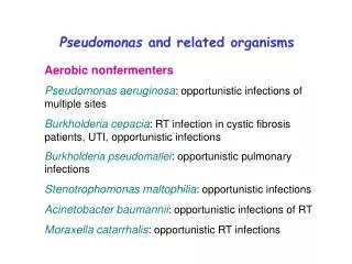
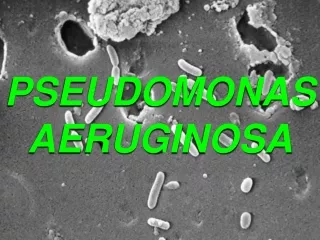
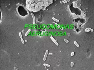
Pseudomonas and related organisms Aerobic gram-negative nonfermentative rods Pseudomonas aeruginosa: opportunistic infections of multiple sites
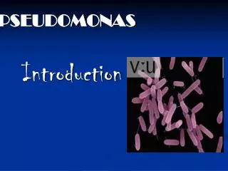
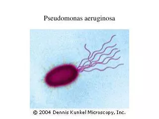
Pseudomonas Structure and Physiology Gram-negative rods. Motile with polar flagella. Obligate aerobe. Oxidase-positive. Do not ferment carbohydrates. Resistant to multiple drugs.
P. aeruginosa Forms round colonies with a fluorescent greenish color, sweet odor, and b-hemolysis. Pyocyanin- nonfluorescent bluish pigment; pyoverdin- fluorescent greenish pigment; pyorubin, andpyomelanin Some strains have a prominent capsule (alginate). Identification of P. aeruginosa is usually based on oxidase test and its colonial morphology: b-hemolysis, the presence of characteristic pigments and sweet odor, and growth at 42 oC.
P. aeruginosa Pathogenesis and Immunity This organism is widely distributed in nature and is commonly present in moist environments in hospitals. It is pathogenic only when introduced into areas devoid of normal defenses, e.g., 1. Disruption of mucous membrane and skin. 2. Usage of intravenous or urinary catheters. 3. Neutropenia (as in cancer therapy). P. aeruginosa can infect almost any external site or organ. P. aeruginosa is invasive and toxigenic. It attaches to and colonizes the mucous membrane or skin, invade locally, and produces systemic diseases and septicemia. P. aeruginosa is resistant to many antibiotics. It becomes dominant when more susceptible bacteria of the normal flora are suppressed.
P. aeruginosa Pathogenesis Antigenic structure, enzymes, and toxins Pili and nonpilus adhesins. Capsule (alginate, glycocalyx): seen in cultures from patients with cystic fibrosis. LPS- endotoxin, multiple immunotypes. Pyocyanin: catalyzes production of toxic forms of oxygen that cause tissue damage. It also induces IL-8 production. Pyoverdin: a siderophore. Proteases Serine protease, metalloprotease andalkaline proteasecause tissue damage and help bacteria spread. Phospholipase C: a hemolysin Exotoxin A: causes tissue necrosis and is lethal for animals (disrupts protein synthesis); immunosuppressive. Exoenzyme S and T: cytotoxic to host cells.
P. aeruginosa Clinical Diseases Infection of wounds and burns (blue-green pus). Patients with severe burns may develop into bacteriemia. Skin and nail infections Meningitis (when introduced by lumbar puncture). Pulmonary infection Tracheobronchitis Necrotizing pneumonia in CF patients: diffuse, bilateral bronchopneumonia with microabscess and necrosis. Eye infections: corneal ulcer. Ear infections Otitis externa: mild in swimmers; malignant (invasive) in diabetic patients. Chronic otitis media Osteochondritisof the foot. Urinary tract infection Sepsis: most cases originate from infections of lower RT, UT, and skin and soft tissue. Ecthyma gangrenosum (hemorrhagic necrosis of skin) may be seen in some patients.
P. aeruginosa Laboratory Diagnosis Specimen: skin lesions, pus, urine, blood, spinal fluid, sputum. Culture: blood agar plate and differential media. Identification of P. aeruginosa is described above. Several subtyping methods, including phage typing and molecular typing, are available for epidemiologic purposes.
Genus Pseudomonas Pseudomonas Pyocyanine Pigment
Genus Pseudomonas Pseudomonas Pyocyanine Pigment
Laboratory diagnosis Nutrient agar Demonstrate pigment production.
Laboratory diagnosis Blood agar spreading flat pigmented, sometime with Beta- hemolysis, mucoid, confluent growth.
Laboratory diagnosis Biochemical tests: Oxidase test, catalase test all are positive. Catalase +ve Oxidase +ve
Combined antibiotic therapy is generally required to avoid resistance that develops rapidly when single drugs are employed. Avoid using inappropriate broad-spectrum antibiotics, which can suppress the normal flora and permit overgrowth of resistant pseudomonads.
P. aeruginosa Prevention and Control Pseudomonas spp. normally inhabit soil, water, and vegetation and can be isolated from the skin, throat, and stool of healthy persons. Spread is via contact with fomites or by ingestion of contaminated food and water. High risk population: patients receiving broad-spectrum antibiotics, with leukemia, burns, cystic fibrosis, and immunosuppression. Methods for control of infection are similar to those for other nosocomial pathogens. Special attention should be paid to sinks, water baths, showers, hot tubs, and other wet areas.
P. aeruginosa Prevention and Control Control: 1. Patients at high risk should not be admitted to a ward where cases of pseudomonas infection are present. 2. Patients infected with P. aeruginosa should be isolated. 3. Sterilize all instruments, apparatus, and dressing; antimicrobial and other therapeutic substances. 4. Monitor clinically relevant isolates of P. aeruginosa by a suitable typing system to identify epidemic strains.