Download

1 / 76
760 likes | 778 Views
Delve into the world of red blood cell disorders, exploring marrow characteristics, anemias, hemolytic conditions, and genetic factors affecting blood health. Learn about clinical manifestations, genetic variations, and triggers for hemolytic anemias, including sickle cell disease and thalassemias.

E N D
RBC and BLEEDING DISORDERS
RBC and Bleeding Disorders • NORMAL • Anatomy, histology • Development • Physiology • ANEMIAS • Blood loss: acute, chronic • Hemolytic • Diminished erythropoesis • POLYCYTHEMIA • BLEEDING DISORDERS
WHERE is MARROW? • Yolk Sac: very early embryo • Liver, Spleen: NEWBORN • BONE • CHILDHOOD: AXIAL SKELETON & APPENDICULAR SKELETON BOTH HAVE RED (active) MARROW • ADULT: AXIAL SKELETON RED MARROW, APPENDICULAR SKELETON YELLOW MARROW
MARROW FEATURES • CELLULARITY 50% • MEGAKARYOCYTES at least 1-2/hpf • M:E RATIO 3:1 • MYELOID MATURATION 1/3 bands or more • ERYTHROID MATURATION nucleus/cytoplasm • LYMPHS, PLASMA CELLS small percentage • STORAGE IRON, i.e., HEMOSIDERIN present • “FOREIGN CELLS”
MARROW “DIFFERENTIATION”
ANEMIAS* • BLOOD LOSS • ACUTE • CHRONIC • IN-creased destruction (HEMOLYTIC) • DE-creased production * A good definition would be a decrease in OXYGEN CARRYING CAPACITY, rather than just a decrease in red blood cells, because you need to have enough blood cells THAT FUNCTION, and not just enough blood cells.
Featuresof ALL anemias • Pallor, where? • Tiredness • Weakness • Dyspnea, why? • Palpitations • Heart Failure (high output), why?
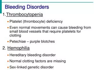
HEMOLYTIC • HEREDITARY • MEMBRANE disorders: e.g., spherocytosis • ENZYME disorders: e.g., G6PD deficciency • HGB disorders (hemoglobinopathies) • ACQUIRED • MEMBRANE disorders (PNH) • ANTIBODY MEDIATED, transfusion or autoantibodies • MECHANICAL TRAUMA • INFECTIONS • DRUGS, TOXINS • HYPERSPLENISM
IMPAIRED PRODUCTION • Disturbance of proliferation and differentiation of stem cells: aplastic anemias, pure RBC aplasia, renal failure • Disturbance of proliferation and maturation of erythroblasts • Defective DNA synthesis: (Megaloblastic) • Defective heme synthesis: (Fe) • Deficient globin synthesis: (Thalassemias)
MODIFIERS • MCV, microcytosis, macrocytosis • MCH • MCHC, hypochromic • RDW, anisocytosis
HEMOLYTIC ANEMIAS • Life span LESS than 120 days • Marrow hyperplasia (M:E), EPO+ • Increased catabolic products, e.g., bilirubin, serum HGB, hemosiderin, haptoglobin-HGB
HEMOLYSIS • INTRA-vascular (vessels) • EXTRA-vascular (spleen)
HEREDITARY SPHEROCYTOSIS Genetic defects affecting ankyrin, spectrin, usually autosomal dominant Children, adults Anemia, hemolysis, jaundice, splenomegaly, gallstones (what kind?)
Glucose-6-Phosphate Dehydrogenase (G6PD) Deficiency • A- and Mediterranean are most significant types
FEATURES of G6PD Defic. • Genetic: Recessive, X-linked • Can be triggered by foods (fava beans), oxidant substances drugs (primaquine, chloroquine), or infections • HGB can precipitate as HEINZ bodies • Acute intravascular hemolysis can occur: • Hemoglobinuria • Hemoglobinemia • Anemia
Sickle Cell Disease • Classic hemoglobinopathy • Normal HGB is α2 β2: β-chain defects (Val->Glu) • Reduced hemoglobin “sickles” in homozygous • 8% of American blacks are heterozygous
Clinical features of HGB-S disease • Severe anemia • Jaundice • PAIN (pain CRISIS) • Vaso-occlusive disease: EVEREWHERE, but clinically significant bone, spleen (autosplenectomy) • Infections: Pneumococcus, Hem. Influ., Salmonella osteomyelitis
THALASSEMIAS • A WIDE VARIETY of diseases involving GLOBIN synthesis, COMPLEX genetics • Alpha or beta chains deficient synthesis involved • Often termed MAJOR or MINOR, depending on severity, silent carriers and “traits” are seen • HEMOLYSIS is uniformly a feature, and microcytic anemia, i.e, LOW MCV (just like iron deficiency anemia has a low MCV) • A “crew cut” skull x-ray appearance may beseen in severe erythroid hyperplasia.
Hemoglobin H Disease • Deletion of THREE alpha chain genes • HGB-H is primarilly Asian • HGB-H has a HIGH affinity for oxygen • HGB-H is unstable and therefore has classical hemolytic behavior
HYDROPS FETALIS • FOUR alpha chain genes are deleted, so this is the MOST SEVERE form of thalassemia • Many/most never make it to term • Children born will have a SEVERE hemolytic anemia as in the erythroblastosis fetalis of Rh disease: • Pallor (as in all anemias), jaundice, kernicterus • Edema (hence the name “hydrops”) • Massive hepatosplenomegaly (hemolysis)
Paroxysmal Nocturnal Hemoglobinuria (PNH) • ACQUIRED, NOT INHERITED like all the previous hemolytic anemias were • ACQUIRED mutations in phosphatidylinositol glycan A (PIGA) • Note: It is “P” and “N” only 25% of the time! GlycosylphosPhatidylInositol (lipid rafts)
Immunohemolytic Anemia • All of these have the presence of antibodies and/or compliment present on RBC surfaces • NOT all are AUTOimmune, some are caused by drugs • Antibodies can be • WARM (IgG) • COLDAGGLUTININ (IgM) • COLD HEMOLYSIN (paroxysmal) (IgG)
IMMUNOHEMOLYTIC ANEMIAS • WARM AGGLUTININS (IgG), will NOT hemolyze at room temp • Primary Idiopathic (most common) • Secondary (Tumors, especially leuk/lymph, drugs) • COLD AGGLUTININS: (IgM), WILL hemolyze at room temp • Mycoplasma pneumoniae, HIV, mononucleosis • COLD HEMOLYSINS: (IgG) Cold Paroxysmal Hemoglobinuria, hemo-LYSIS in body, ALSO often follows mycoplasma pneumoniae
COOMBSTEST • DIRECT: Patient’s CELLS are tested for surface Ab’s • INDIRECT: Patient’s SERUM is tested for Ab’s.
HEMOLYSIS/HEMOLYTIC ANEMIAS DUE TO RBC TRAUMA • Mechanical heart valves breaking RBC’s • MICROANGIOPATHIES: • TTP • Hemolytic Uremic Syndrome
NON-Hemolytic Anemias:i.e., DE-creased Production • “Megaloblastic” Anemias • B12 Deficiency (Pernicious Anemia) • Folate Deficiency • Iron Deficiency • Anemia of Chronic Disease • Aplastic Anemia • “Pure” Red Cell Aplasia • OTHER forms of Marrow Failure
MEGALOBLASTIC ANEMIAS • Differentiating megaloblasts (marrow) from macrocytes (peripheral smear, MCV>94) • Impaired DNA synthesis • For all practical purposes, also called the anemias of B12 and FOLATE deficiency • Often VERY hyperplastic/hypercellular marrow
Vit-B12 Physiology • Oral ingestion • Combines with INTRINSIC FACTOR in the gastric mucosa • Absorbed in the terminal ileum • DEFECTS at ANY of these sites can produce a MEGALOBLASTIC anemia
Please remember that ALL megaloblastic anemias are also MACROCYTIC (MCV>94 or MCV~100), and that not only are the RBC’s BIG and hyperplastic/hypercellular, but so are the neutrophils, and neutrophilic precursors in the bone marrow too, and even more so, HYPERSEGMENTED!!!
PERNICIOUS ANEMIA • MEGALOBLASTIC anemia • LEUKOPENIA and HYPERSEGS • JAUNDICE • NEUROLOGIC posterolateral spinal tracts • ACHLORHYDRIA • Can’t absorb B12 • LOW serum B12 • Flunk Schilling test, i.e., can’t absorb B12, using a radioactive tracer
FOLATE DEFICIENCY MEGALOBLASTIC AMEMIAS • Decreased Intake: diet, etoh-ism, infancy • Impaired Absorption: intestinal disease • DRUGS: anticonvulsants, BCPs, CHEMO • Increased Loss: Hemodialysis • Increased Requirement: Pregnancy, infancy • Impaired Usage
Fe Deficiency Anemia • Due to increased loss or decreased ingestion, almost always, in USA, nowadays, increased loss is the reason • Microcytic (low MCV), Hypochromic (low MCHC) • THE ONLY WAY WE CAN LOSE IRON IS BY LOSING BLOOD, because FE is recycled!
Fe Transferrin Ferritin (GREAT test) Hemosiderin
Clinical Fe-Defic-Anemia • Adult men: GI Blood Loss • PRE menopausal women: menorrhagia • POST menopausal women: GI Blood Loss
2 BEST lab tests: • Serum Ferritin • Prussian blue hemosiderin stain of marrow (also called an “iron” stain)
Anemia of Chronic Disease* • CHRONIC INFECTIONS • CHRONIC IMMUNE DISORDERS • NEOPLASMS • LIVER, KIDNEY failure * Please remember these patients may very very much look like iron deficiency anemia, BUT, they have ABUNDANT STAINABLE HEMOSIDERIN in the marrow!
APLASTIC ANEMIAS • ALMOST ALWAYS involve platelet and WBC suppression as well • Some are idiopathic, but MOST are related to drugs, radiation • FANCONI’s ANEMIA is the only one that is inherited, and NOT acquired • Act at STEM CELL level, except for “pure” red cell aplasia