
Cell Structure and Function: Discovery and Microscope Tools
E N D
Presentation Transcript
CHAPTER THREE CELL STRUCTURE AND FUNCTION
CONTRIBUTORS TO THE DISCOVERY OF CELLS HOOKE LEEUWENHOEK _____________ SCHLEIDEN SCHWANN VIRCHOW _____________ _____________ _____________ _____________ RESPONSIBLE FOR CELL THEORY CORK ANIMALCULES Made better ______________ and observed cells in greater ______________. First to observe ______________ Concluded that all ___________ ___________ were made up of ___________ Proposed that all cells come from ____________ __________ The first to __________ cells. Responsible for ____________ them The first to note that _____________ were made up of ___________
CELL THEORY • ___________________________________________________________ • 2. ___________________________________________________________ • 3. ___________________________________________________________
TOOLS OF BIOLOGY • Microscope Function Magnifies up to… • ______________ microscope Uses light. __________ • ______________ microscope Light cannot pass. _________ • ______________ microscope Uses electrons __________ COMPOUND LIGHT 1000X STEREO 40X Also known as ______________ scope DISSECTING 500,000X ELECTRON COMPOUND LIGHT STEREOSCOPE ELECTRON MICROSCOPE
LIGHT MICROSCOPE eyepiece Body Tube Turrett Arm Focus Objective Low Objective Stage High Power Objective Stage Clips Course Adjustment Diaphragm Fine Adjustment Light Source Base
Labeling the Parts of the Microscope (Independent Practice) EYEPIECE 1. 2. BODYTUBE TURRETT 3. ARM 10. 11. FOCUS OBJECTIVE 4. LOW POWER OBJECTIVE 5. STAGE CLIPS 12. HIGH POWER OBJECTIVE 13. COURSE ADJUSTMENT 6. STAGE 7. DIAPHRAGM 8. LIGHT SOURCE 14. FINE ADJUSTMENT 9. BASE
TOTAL MAGNIFICATION The focus objective focuses __________ The low power objective focuses _______ The high power objective focuses _______ 4X 10X 40X Keep in mind, there is also a lens in the EYEPIECE that focuses __________ “ON TOP OF” the magnification of the objective lenses. Therefore, _____________________________would be: _______________ X _________________ 10X TOTAL MAGNIFICATION OBJECTIVE EYEPIECE Practice: EYEPIECE X OBJECTIVE = TOTAL MAGNIFICATION TOTAL MAGNIFICATION OF FOCUS POWER __________ X __________ = ______________ TOTAL MAGNIFICATION OF LOW POWER __________ X __________ = ______________ TOTAL MAGNIFICATION OF HIGH POWER __________ X __________ = ______________ 10 4 40 X 100 X 10 X 10 X 10 X 40 X 400 X
EYEPIECE: BODY TUBE: Where you place your eye. Contains ______ ______ that usually magnifies ______. Tube that supports the ______ _______ and connects it to the _________________. EYE PIECE ONE LENS TURRETT/NOSE PIECE 10x
OBJECTIVES: STAGE CLIPS: ______________ that magnify objects to varying __________. FOCUS OBJECTIVE: ______________________________________________ LOW POWER OBJECTIVE: ______________________________________________ HIGH POWER OBJECTIVE: ______________________________________________ LENSES Holds the _____________ in place SLIDE “POWERS” SHORTEST LENS (4X) ONLY USED FOR SCANNING SMALL LENS (10 X) LOW MAGNIFYING POWER LONGEST LENS (40 X) HIGH MAGNIFYING POWER
ADJUSTMENTS BASE Knobs that make adjustments to the ______________ COURSE ADJUSTMENT ___________________________________________________________________________ FINE ADJUSTMENT ___________________________________________________________________________ FOCUS Supports the _____________ MICROSCOPE MAKES LARGE ADJUSTMENTS USED WITH FOCUS AND LOW POWER OBJECTIVES MAKES SMALL ADJUSTMENTS USED WITH HIGH POWER OBJECTIVE ONLY
LIGHT SOURCE: Directs light up through the ______________ and through the ______________ so that it may be ______________ DIAPHRAGM SPECIMEN VIEWED
STAGE: Supports the __________________ SLIDE/SPECIMEN
NOSEPIECE: Also known as the _______________. It is the rotating device that holds the _____________/ (_________). TURRETT LENSES OBJECTIVES
DIAPHRAGM An adjustable ________________ under the stage, allowing different __________ of __________ onto the stage. OPENING AMOUNTS LIGHT
____________________________________________________________________________________________________________________________ arm - this attaches the eyepiece and body tube to the base.base - this supports the microscope.body tube - the tube that supports the eyepiece.coarse focus adjustment - a knob that makes large adjustments to the focus.diaphragm - an adjustable opening under the stage, allowing different amounts of light onto the stage.eyepiece - where you place your eye.fine focus adjustment - a knob that makes small adjustments to the focus (it is often smaller than the coarse focus knob).high-power objective - a large lens with high magnifying power.inclination joint - an adjustable joint that lets the arm tilt at various angles.low-power objective - a small lens with low magnifying power.mirror (or light source) - this directs light upwards onto the slide.revolving nosepiece - the rotating device that holds the objectives (lenses).stage - the platform on which a slide is placed.stage clips - metal clips that hold a slide securely onto the stage. _____________________________________________________________ __________________________________________________________ ____________________________________________________ ____________________________________________________ ________________________________________________________ ____________________________________________________ ____________________________________________________ ____________________________________________________ ____________________________________________________ ____________________________________________________ __________________________________________________________ ____________________________________________________
arm - this attaches the eyepiece and body tube to the base.base - this supports the microscope.body tube - the tube that supports the eyepiece.coarse focus adjustment - a knob that makes large adjustments to the focus.diaphragm - an adjustable opening under the stage, allowing different amounts of light onto the stage.eyepiece - where you place your eye.fine focus adjustment - a knob that makes small adjustments to the focus (it is often smaller than the coarse focus knob).high-power objective - a large lens with high magnifying power.inclination joint - an adjustable joint that lets the arm tilt at various angles.low-power objective - a small lens with low magnifying power.mirror (or light source) - this directs light upwards onto the slide.revolving nosepiece - the rotating device that holds the objectives (lenses).stage - the platform on which a slide is placed.stage clips - metal clips that hold a slide securely onto the stage.
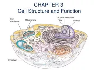
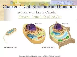
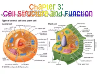
Types of cells • Prokaryotic cells • primitive cells – “before the nucleus” • No membrane bound organelles • No nucleus • Found in Eubacteria and Archaeabacteria • Cell wall, cell membrane, cytoplasm, DNA & ribosomes
Eukaryotic “true nucleus” • Have membrane – bound organelles • All other kingdoms but Archaeabacteria & Eubacteria • Organelles such as: Mitochondria, endoplasmic reticulum, vacuoles, lysosomes, and chloroplasts.
Structures common to both Prokaryotic and Eukaryotic cells • Cell Membrane • Ribosomes • DNA • Cytoplasm • Cell Walls(not in Animal or some Protist cells though!) • Organelle – small structures in cells that have a specific function
ANIMAL CELL PLANT CELL http://www.johnkyrk.com/CellIndex.html
CELL MEMBRANE __________________________________________________________________________________________________ __________________________________________________________________________________________________ CELL MEMBRANE _______________ _______________
CYTOPLASM ________________________________________________________________________________________ ____________________________________________ CYTOPLASM _______________
NUCLEUS ___________________________________________________ _________________________________________________ __________________________________ __________________________________ __________________________________ __________________________________ _______________
NUCLEAR MEMBRANE _________________________________________ Nuclear Membrane in Green ______________________________ ______________________________
CHROMOSOMES ____________________________________________________________________________________ Chromosomes in Red _______________
NUCLEOLUS _____________________________________ _______________
MITOCHONDRIA __________________________________________________________________________ ____________
LYSOSOMES ________________________________________________________________________________________ ____________
GOLGI APPARATUS _______________________________________ ______________ ______________
ENDOPLASMIC RETICULUM ________________________________________________________________________________________ ____________________________________________ _______________ _______________
RIBOSOMES __________________________________________________________________________________________________________________________________________________________________ _______________
CENTRIOLES ____________________________________________
VACUOLE _______________________________________ ANIMAL CELL PLANT CELL _______________________________________________________________________________________ ____________________________ ________ VACUOLE
CELL WALL _________________________________________________________ __________________________________________________________ __________________________________________________________ ____________
Cytoskeleton • A network of protein filaments that help the cell to maintain its shape. • Centrioles – microtubules in animal cells that are involved in moving things during cell division. • Also involved in • movement.
CHLOROPLAST ____________________________________________________________________________________________________________________________________ ______________________ CHLOROPLASTS
ANIMAL CELL DIAGRAM Label the organelles of this animal cell. • _________________ • _________________ • _________________ • _________________ • _________________ • _________________ • _________________ • _________________ • _________________ • _________________ • _________________ • _________________ • _________________
2.__________________ 1.__________________ 12.__________________ 3.__________________ 11.__________________ 4.__________________ 10.__________________ 5.__________________ 9.__________________ 8.__________________ 7.__________________ 6.__________________
Structure of Lipid Bilayer Section 3 Cell Organelles and Features Chapter 3
Section 3 Cell Organelles and Features Chapter 4 Plasma Membrane, continued • Membrane Proteins • Cell membranes often contain proteins embedded within the phospholipid bilayer.-called integral prot
Diffusion Section 3 Passive Transport Chapter 5 • Passive transport– Involves the movement of molecules across the cell membrane without an input of energy from the cell.es the movement of molecules across cell membrane without an input of rDiffusion is the movement of molecules from an area of higher concentration to an area of lower concentration, driven by the molecules’ kinetic energy until equilibrium is reached. • gy by the cell. • is the movement of molecules from an area of higher concentration to an area of lower concentration, driven by the molecules’ kinetic energy until equilibrium is reached.
Diffusion • Concentration Gradient - The difference in the concentration of molecules across a distance. • Equilibrium - When the concentration of molecules is the same throughout the space the molecules occupy. • Once equilibrium is reached, molecules move randomely.
Diffusion, continued Section 3 Passive Transport Chapter 5 • Diffusion Across Membranes • Molecules can diffuse across a cell membrane by dissolving in the phospholipid bilayer or by passing through pores in the membrane. • Depends on size and type of molecule and on the chemical nature of the membrane.
Diffusion Section 3 Passive Transport Chapter 5
Osmosis Section 3 Passive Transport Chapter 5 • Osmosisis the diffusion of water across a membrane. • No energy is required (passive transport).
Osmosis, continued Section 3 Passive Transport Chapter 5 • Direction of Osmosis • The net direction of osmosis is determined by the relative solute concentrations on the two sides of the membrane.
Osmosis, continued Section 3 Passive Transport Chapter 5 • Direction of Osmosis • When the solute concentration outside the cell is higher than that in the cytosol, the solution outside is hypertonic to the cytosol, and water will diffuse out of the cell.
Osmosis, continued Section 3Passive Transport Chapter 5 • Direction of Osmosis • When the solute concentrations outside and inside the cell are equal, the solution outside is isotonic, and there will be no net movement of water.