Download
1 / 28
280 likes | 439 Views
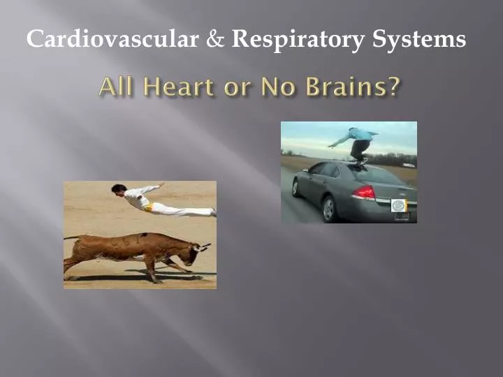
All Heart or No Brains?. Cardiovascular & Respiratory Systems. Question: How much does a Normal Heart Weigh?. 5 ounces 11 ounces About 1 pound 2.5 pounds. B. 11 Ounces on average. ( The average brain weighs 3 pounds or 48 ounces). Statistics. Beats per Minute = 60-80

E N D
All Heart or No Brains? Cardiovascular & Respiratory Systems
Question: How much does a Normal Heart Weigh? 5 ounces 11 ounces About 1 pound 2.5 pounds B. 11Ounces on average. (The average brain weighs 3 pounds or 48 ounces)
Statistics Beats per Minute = 60-80 Beats per Day = 100,000 Beats per Year = 35 Million Beats per Lifetime = 2.5 Billion *Blood Pumped per minute (Rest) = 5 Liters/ 5 Quarts Blood Pumped to over 75 Trillion Cells in Body Through over 60,000 miles of vessels
*Cardiovascular System Cardio – pertaining to Heart Vascular- = a little vessel, consisting of cells joined into tubes for transporting/circulating CV system – Body system transports nutrients, gases, hormones, cellular waste products.
*Major Cardiovascular Functions wDelivery:. oxygen and nutrients wRemoval:carbon dioxide and waste products wTransportation: hormones wMaintenance: body temperature, pH wPrevention: infection (Helps Immune System function)
Origin of Heart Shape Heart is Center of All Emotions. Heavy Heart 7th century B.C. city-state of Cyrene. Cyrene traded the rare, now extinct, plant silphium. It was known as a means for birth control. The seedpod of the silphium looks exactly like a valentine's heart. Moreover, its use in sex is an obvious connection to love
The Circulatory System • Heart • Pumps blood • Arteries and arterioles • Carry blood away from the heart • Capillaries • Exchange of nutrients with tissues • Veins and venules • Carry blood toward the heart
The Basics Thump-thump?
Blood Vessels • Artery - a high-pressure vessel, possess a thick layer of smooth muscle to help off set the high blood pressures it must endure, can constrict or dilate (Major Arteries) • Arteriole- a resistance vessel, possess a circular smooth muscle layer, is the primary site of blood flow regulation • Capillary – site of nutrient transfer (Diffusion) • Venule– a pre-vein, small amount of muscle • Vein – a capacitance vessel, very elastic, capable of “holding” blood; posses valves to prevent backflow
The Skeletal Muscle Pump • Rhythmic skeletal muscle contractions force blood in the extremities toward the heart • One-way valves in veins prevent backflow of blood
Pulmonary & Systemic Circuits Respiratory System VR-RA-RV-Lungs-LA-LV-Aorta-Body Coronary Arteries-Heart
*Aorta The Main artery & Largest (elastic). When the left ventricle contracts to force blood into the aorta, the aorta expands. This stretching gives the potential energy that will help maintain blood pressure during diastole as during this time the aorta contracts passively.
Left Ventricle • The left ventricle receives oxygenated blood from the left atrium via the bicuspid valve. The blood is then transported throughout the body. • The thick walls of the left ventricle pumps blood to the aorta, the systemic arteries, the capillaries, the veins, and back to the right atrium. • The left ventricle is also the main function of stroke volume and cardiac output.
Cardiac Output Graph (11) Muscles receive most of the cardiac output during exercise because the muscles are the ones doing most of the work!
Heart Wall Anatomy • Epicardium– a connective tissue covering of the heart which contains blood vessels, lymph vessels, and nerve endings; provides a lubrication buffer for the heart • Myocardium– the cardiac muscle layer; the myocardium will be discussed in greater detail in the next slide • Endocardium – the inner layer of endothelial cells and more collagen (connective) tissues
Myocardium—The Cardiac Muscle w Thickness varies directly with stress placed on chamber walls. w Left ventricle is the most powerful of chambers and thus, the largest. w With vigorous exercise, the left ventricle size increases. • The heart has autoconduction, i.e., it beats on its own without nerve or hormonal control. Due to intercalated disks—impulses travel quickly in cardiac muscle and allow it to act as one large muscle fiber; all fibers contract together. Fig 9.3
Myocardium • Myocardium is striated muscle tissue, so it has some similar aspects to skeletal muscle • However, it also has several qualities which are much different
Myocardium cont. • Differences from skeletal muscle: • *Only one fiber type, so there are no fiber-type differences within the myocardium • Cardiac muscle fibers are relatively short. Most are only a few hundred sarcomeres in length • *Cardiac muscle is not voluntary muscle • Cardiac muscle fibers are connected by intercalated disc; creates a functional syncytium
Electrocardiogram • *Records the electrical activity of the heart • P-wave • Atrial depolarization • QRS complex • Ventricular depolarization • T-wave • Ventricular repolarization
Review • Video
Breathing Mechanics http://youtu.be/hp-gCvW8PRY • Normal Inspiration: Diaphragm contracts (shortens) creating a vacuum in the chest cavity. • Normal Expiration: Diaphragm relaxes and the chest cavity collapses forcing air out. • Exercising Inspiration: Diaphragm, intercostal, and abdominal muscles contract to lift the rib cage and create a greater vacuum in the chest cavity. • Exercising Expiration: All muscle relax and the chest cavity collapses forcing air out.