Download
1 / 21
210 likes | 468 Views
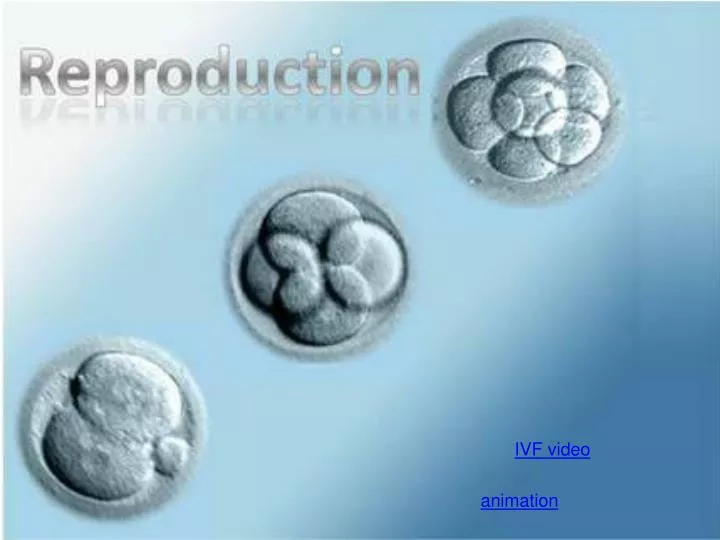
IVF video. animation. 11.4.1 Annotate a light micrograph of testis tissue to show the location and function of interstitial cells (Leydig cells), germinal epithelium cells, developing spermatozoa and Sertoli cells. Testis Structure. Vas Deferens. Epididymus. Seminiferous Tubules. Scrotum.

E N D
IVF video animation
11.4.1 Annotate a light micrograph of testis tissue to show the location and function of interstitial cells (Leydig cells), germinal epithelium cells, developing spermatozoa and Sertoli cells. Testis Structure Vas Deferens Epididymus Seminiferous Tubules Scrotum Testis animation Tunica Albuguinea
Sertoli cells spermatids Interstitial Cells Spermatogonia Interstitial Cells
11.4.2 Outline the processes involved in spermatogenesis within the testis, including mitosis, cell growth, the two divisions of meiosis and cell differentiation. Spermatogenesis Seminiferous Tubule lumen Developing Sperm Sertoli Cell Leydig Cells (Interstitial Cells)
11.4.3 State the role of LH, testosterone and FSH in spermatogensis. Spermatogonium (germinal epithelium) Primary Spermatocyte Secondary Spermatocyte Spermatids Nourishment from Sertoli cells Spermatozoa (mature sperm cells)
11.4.3 State the role of LH, testosterone and FSH in spermatogensis. FSH is produced an released by the anterior pituitary gland and stimulates meiosis of the primary spermatocytes, giving secondary spermatocytes (now haploid). LH is also released and reaches the interstitial cells (Leydig cells) of the testis, where it stimulates testosterone production. Testosterone stimulates the final meiotic division and differentiation of the spermatids to form mature spermatozoa.
11.4.7 Outline the role of the epididymis, seminal vesicle and prostate gland in the production of semen. Epididymus Seminal vesicle Prostate gland
11.4.6 Draw and label a diagram of a mature sperm and egg. acrosome flagellum mitochondria
11.4.3 Annotate a diagram of the ovary to show the location and function of germinal epithelium, primary follicles, mature follicle and secondary oocyte. Primary oocyte Primary follicle Germinal epithelium Secondary oocyte Zona pellucida Corpus luteum
Germinal epithelium Primary follicle mature follicle Secondary oocyte Zona pellucida medulla
11.4.5 Outline the processes involved in oogenesis within the ovary, including mitosis, cell growth, the two divisions of meiosis, the unequal division of cytoplasm and the degeneration of polar body.
11.4.8 compare the processes of spermatogenesis and oogenesis, including the number of gametes and the timing of the formation and release of gametes. mitosis LH and FSH 2 meiotic divisions
11.4.9 Describe the process of fertilization, including the acrosome reaction, penetration of the egg membrane by a sperm and the cortical reaction.
11.4.9 Describe the process of fertilization, including the acrosome reaction, penetration of the egg membrane by a sperm and the cortical reaction.
11.4.9 Describe the process of fertilization, including the acrosome reaction, penetration of the egg membrane by a sperm and the cortical reaction.
11.4.10 Outline the role of HCG in early pregnancy. Progesterone Estrogen HCG • HCG: • Secretion of Progesterone and Estrogen by Corpus Luteum • Progesterone and Estrogen inhibit FSH-LH – so no more eggs are released • Progesterone maintain endometrium, where the blastocyst develops into • a fetus and the placenta forms
11.4.12 Explain how the structure and functions of the placenta, including its hormonal role in secretion of estrogen and progesterone, maintain pregnancy. 11.4.13 State that the fetus is supported and protected by the amniotic sac and amniotic fluid. 11.4.14 State that the materials are exchanged Between the maternal and fetal blood In the placenta. • Functions of the placenta • Releases _____________ and • _________ • Exchange of materials between • _________ and ________ blood • (blood does not mix) progesterone estrogen fetal maternal
11.4.12 Explain how the structure and functions of the placenta, including its hormonal role in secretion of estrogen and progesterone, maintain pregnancy. • Rich fetal blood supply • Few thin membranes give a short • diffusion path for materials. • Villi increase surface area for • exchange • Placenta has a large surface area • and many RER and vesicles for • production of hormones
11.4.15 Outline the process of birth and its hormonal control, including the changes in progesterone and oxytocin levels and positive feedback. oxytocin contrations further release of oxytocin
11.4.15 Outline the process of birth and its hormonal control, including the changes in progesterone and oxytocin levels and positive feedback. Cervix is dialating placenta