Cardiovascular System
240 likes | 281 Views
Dive into the intricate functions and physiology of the heart - from generating blood pressure to regulating blood flow. Explore the cardiac cycle, heart conduction, and the importance of cardiac output.

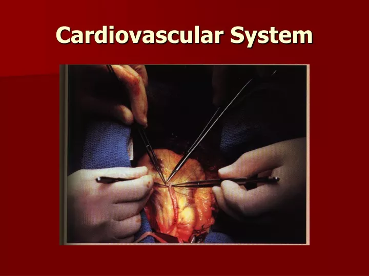
Cardiovascular System
E N D
Presentation Transcript
I. Functions of the heart. 1. Generating blood pressure. 2. Routing blood. 3. Ensuring blood moves one way. 4. Regulating blood supply.
Cardiovascular System II. Heart A. General Characteristics • 1. 100,000 time/day • 2. 4,00L/day = 1,000 gallons • 3. 60,000 miles of blood vessels • 4. Center of the circulatory System • 5. Center of the Thoracic Cavity, between the lungs • 6. 2/3 on the left side • 7. Decreases in size after the age of 65.
B. Coverings of the heart 1. Pericardium - covers the heart, 2 main layers a. Parietal Cardium (pericardial sac) •Outer •Loose fitting 1. Fibrous - Outer, thick, tough dense connective tissue. Protects & anchors to the diaphragm 2. Serous - Inner, thin, Squamous epithelium b. Visceral Pericardium (Epicardium) •Attaches to the surface of the heart •Considered to be the outer most layer of the heart. c. Pericardial Cavity •Space filled with fluid •Lubrication •Percarditis - swelling of space
C. Heart Walls 3 layers - middle is the most important - Myocardium • Epicardium - Thin protective barrier of the heart Serous membrane Fat deposits 2. Myocardium -Bulk of the heart Fibrous skeleton - bundles of connective tissue Layer that contracts 3. Endocardium- Inner most layer Smooth white layer • Squamous epithelium
D. Heart Chambers Superior • Atria - Receiving chambers for blood Min. role in pumping Pectinate Muscles - ridges of muscle Interatrial Septum - separates the atrium into right & left halves FossalOvalis - oval depression of the heart believed to be an opening in the fetal heart a. Right Atrium - receives from the vena cava b. Left Atrium receives from the lungs
Inferior 2. Ventricles - forces blood through the body Trabeculae Carnae - Irregular folds of muscle in the endocardium Papillary muscles - Slender projections off of the trabuclae carnae • Attaches to valves & aids in function Interventricular Septum - Separates ventricles into left & right halves Coronary Sulcus - Externally separates the atrium & ventricles
E. Heart valves Blood flows in one direction Prevents back flow • 1. Atrioventricular valves (AV) - between atrium & ventricles 2 or 3 triangular flaps or cusps Point downward into the ventricles Tricuspid between right Atrium & ventricle (3 flaps) Bicuspid (mitral) between left atrium & ventricle (2 flaps) Chordae tendinae - strands of connective tissue Anchors cusps to papillary walls of the ventricles Murmur - Cusps do not lose completely - leaking of blood
2. Semilunar (SL) Valves Between ventricles & blood leaving Pulmonary & Aortic 3 half moon (semilunar) cusps
G. Supply of blood to the heart Coronary Circulation Right & Left Coronary Arteries - carries fresh oxygenated blood (70% of oxygen, only 25% to skeletal muscles, increases to 70% during exercise) Great & Small cardiac vein Coronary Sinus - large vein that collects blood leaving the heart
II. Heart Physiology - Pumps blood through out the body A. Cardiac Cycle - contraction of both the atria & then ventricle. 1. Systole - Contraction 2. Diastole - Relaxation B. Heart Sounds - LUB - DUB Closing of the heart valves 1. Lub - Closing of the AV valves 2. Dub - Closing of the Sl valves
C. Heart Conduction - Each cardiac cycle is stimulated by special conducting cells in the heart. 1. Receives a signal form the autonomic nervous system. 2. Sinoatrial (SA) Node “Pacemaker” - cluster of pace setting cells Initiates each cardiac cycle by generating an electric impulse. Spread quickly through out the atrium. Stimulates the second cluster of cells.
3. Atrioventricular (AV) Node, AV. bundle, Bundle of His - relays the signal to the ventricles. Extends down the septum of the heart. 4. Purkinje fibers - branches of the AV node, passes further into the myocardium. 5. If the SA node is unable to produce the electrical impulse for the heart to contract, the AV node functions as the pacemaker 6. Slower – Ectopic beat.
D. Electrocardiogram (ECG or EKG) - measures the electrical events during a cardiac cycle 1. Detect changes in the electrical changes in the heart wall 2. Electrical changes produces a changes in the ionic flow through out the body SA node fires send action potential. P Wave - depolarization of the atria - action potential. QRS Wave - depolarization of the ventricle. T Wave ventricular repolarization of the ventricle
PQ interval – atria contract & begins to relax. QT interval – ventricle depolarizes & repolarizes.
E. Cardiac Output - Volume of blood pumped 1. Heart rate X Stroke Volume = Cardiac Output 75 bpm X 70 ml = 5250 ml/min (5.25 L/min) 2. Adjustments - exercise Heart rate Stroke Volume 3. Starling’s Law - Further the heart is stretched, the stronger the contraction. Preload – pressure on the heart when the ventricles are stretched when filling with blood. Afterload – pressure the heart must beat against.
F. Regulation of Heart Activity 1. Controlled by the reflex center (cardioregulatory center) - medulla oblongata. 2. Baroreceptors - detect the blood pressure. 3. Parasympathetic fibers from the medulla oblongata through the Vagus nerve extends to the heart. Acetylcholine (Ach) slows the heart Norepinephrine (NE) speed the heart up
G. Cardiac cycle Three major events in the cycle. 1. Systole – a. Blood is pushed towards the atria, closely the AV valves. b. Pressure increases in the ventricle forcing the SL valves to open. 2. Diastole – a. Pressure in the ventricles decrease, the AV valves open and blood fills the ventricles up to 70% of their volume. 3. End- Atria relaxes & fills with blood, then contract & starts it over.
III. Aging A. By 70 output is reduced by 30%, by 85 30%-60%. B. Hypertrophy is common (enlargement of left ventricle), due to increase afterload (high blood pressure). Leads to decreased elasticity & increased stiffness. Increased left atria pressure and cause pulmonary edema, feel out of breath. C. Greater amount of time to contract & relax leading to decreased in maxmium heart rate. D. Connective tissue with the valves becomes less flexible. E. Development of coronary artery disease in 10% of people over 80.
VI. Cardiovascular diseases • Congestive heart failure - failure of the heart to pump blood to the body tissues. B. Heart Block – Failure of the SA or AV. to generate impulses. C. Heart fibrillation - Heart beats at a irregular pace. D. Heart flutter - heart race up to 300 bpm. E. Hypertension - elevated blood pressure. F. Murmur - Leaking of blood through a closed valve. G. Myocarditis - infection of the heart muscle. H. Pericarditis - Infection of the pericardial sac which results in thicken or scarring.
1. What are the three types of blood vessels and how are they different? 2. What is the difference between vasodialation & vasoconstriction? 3. What are the 3 layers of the blood vessels? 4. Does blood ever flow in reverse within the blood vessels?