Download
1 / 25
250 likes | 275 Views
Learn the basics of ultrasound knobology and how to use the machine effectively. Explore core applications like central lines, early pregnancy ultrasound, gallbladder examination, and renal imaging. Get insights on credentialing requirements and legal aspects.

E N D
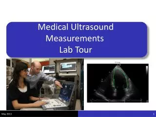
Ultrasound : Zonare Knobology Jamie Jenkins MD, RDMS Regional Ultrasound Director FHS St Josephs Medical Center
Knobology How to use the machine
Medical Legal Aspect • Only 21% of EM physicians personally perform bedside ultrasound • Standard of Care: Central lines • No lawsuits for misreads as of 2007 • 3 lawsuits for failure to use • 2 with AAA • 1 with rule out ectopic • Credentialing • Can we hide behind it or with out it?
Changing probes • Press the transducer key and then use the soft keys to select which transducer you need • Please do not change the Transducer plug-ins.
New patient exam Press new patient key in upper left hand corner of the keyboard
New Patient: Non-credentialed physicians, Training scans at all hospitals • Last name: Training • First name: EDUS • Do not fill out MR number • Operator: Your name • Press exit • On first page, press space bar and type in pt name and MR number
New Patient: Credentialed physicians At St Joes & St Francis • Enter an order in EPIC • Click worklist • Select patients name from list • The screen will self populate • Fill in your name under operator • Press exit and start scanning
New Patient: Credentialed physicians at St Anthony & St Clare • Last name: Patients last name • First name: Patients first name • ID: MR number • Operator: Your last name • Press exit and start scanning • Print images and attach to a paper with pts sticker and give to HUC to scan in for HIM
If you manually enter info instead of using worklist: St Josephs & St Francis • If you manually enter the patient information including the MR number • No leading zeros • No dashes • No FIN numbers • Ex: MR number is 001-234-567 • Enter 1234567 • At St Josephs and St Francis you still need to enter an order after you finish the scan • It is preferred that you use the worklist
Central Lines • Document on central line section of the note • Wire or needle seen in lumen • Image saved to patients permanent record • You must save an image of this. • There is an EPIC smart Phrase under my name (Jamie Jenkins) .eduscentralline
OB/ IUP • OB/ Pregnancy Scan • OB mode • Calc button will give you options for fetal dates/ FHT • Transabdominal views of the uterus • transverse and longitudinal • Fetal Heart Tones (m-mode) • Fetal dates • >12weeks BPD • <12weeks crown rump length • BPD (Inner to outer walls of the skull) • Clips of bilateral adenexa (even if obvious IUP)
Fetal Heart Tones • Please use M –mode to document FHT. • Press M-mode once and the screen will change, • Align the line with the fetal heart • When you have the image you want press freeze • Press calc, select FHT/ HR and measure the distance between beats.
Early Pregnancy Ultrasound • Misconception: A very low B-HCG rules out ectopic pregnancy • Truth: 40% of ectopic pregnancies will present with a B-hcg less than 1000 mIU/ml • 20% will present with a B-hcg less than 500 mIU/ml • Pts who present with a B-hcg less than 1000 mIU/ml have a higher risk of ectopic pregnancy • Keep in mind with ectopics all you may see is an empty uterus and free fluid in the pelvis.
Gallbladder • Transverse and longitudinal clips through the gallbladder • Measurement of the anterior wall of the gallbladder • (2-3 mm) • Measurement of the CBD • (4-8mm) • Look for: • Stones • Wall thickness • Pericholecystic fluid • Sono-murphys • CBD dilation
Gallbladder • Common Bile Duct
Gallbladder Literature • Indications: Signs and symptoms of Cholecystitis • Most sensitive signs: Stones and sono-murphys sign • Very specific signs: Wall thickness, pericholecystic fluid
FAST • Clips of • RUQ • LUQ • Suprapubic • Cardiac • Depth: enough to see posterior surface of heart • Pericardial vs Pleural effusion • Parasternal long if unable to get Subxiphoid view
Aorta • Transverse View • epigastric and infra-renal • Measure the outer to outer wall of the Aorta • Nl <3cm • longitudinal views of the aorta in the epigastric region • Bifurcation of the iliacs
Renal • Bilateral Kidney’s • Two views of each: transverse and longitudinal • Bladder (2 views) • Please label your views
Credentialing • 50 scans to remain credentialed in all areas of ultrasound over 2 years • CME is required as well • This lecture • Scanning shifts with me • Ultrasound courses/lectures at conferences
Questions?????? • If there are any questions please feel free to email me. • Jamiegoodis@gmail.com