
Inflammation: Process, Repair, and Response
Learn about the cardinal signs of inflammation, vascular responses, cell reactions, repair mechanisms, and the interplay between inflammation and repair. Acute and chronic patterns, cellular components, chemical mediators, and termination processes are also explored in this comprehensive guide.

Inflammation: Process, Repair, and Response
E N D
Presentation Transcript
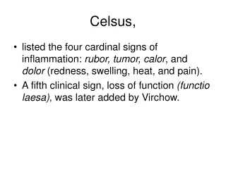
Celsus, • listed the four cardinal signs of inflammation: rubor, tumor, calor, and dolor (redness, swelling, heat, and pain). • A fifth clinical sign, loss of function (functio laesa), was later added by Virchow.
Scottish surgeon John Hunter • inflammation is not a disease but a nonspecific response • Julius Cohnheim (1839-1884) first used the microscope to observe inflamed blood vessels in thin, transparent membranes, such as in the mesentery and tongue of the frog. • Noted the initial changes in blood flow, the subsequent edema caused by increased vascular permeability, and the characteristic leukocyte emigration,
the Russian biologist Elie Metchnikoff 1880s, • discovered the process of phagocytosis by observing eating bacteria by mammalian leukocytes. • concluded the purpose of inflammation was to bring phagocytic cells to the injured area to engulf invading bacteria. • both cells (phagocytes) and serum factors (antibodies) were critical for defense against microorganisms,
Sir Thomas Lewis, • that chemical substances, such as histamine locally induced by injury, mediate the vascular changes of inflammation.
Definition • inflammatory process is the reaction of blood vessels, leading to the accumulation of fluid and leukocytes in extravascular tissues. • Inflammation is a complex reaction to injurious agents such as microbes and damaged, usually necrotic, cells that consists of vascular responses, migration and activation of leukocytes, and systemic reactions.
Definition • a host response of vascularized tissues to exogenous and endogenous stimuli is called inflammation.
The inflammatory response is closely intertwined with the process of repair. • Inflammation serves to destroy, dilute, or wall off the injurious agent, and it sets into motion a series of events that try to heal and reconstitute the damaged tissue.
Repair • Repair begins during the early phases of inflammation but reaches completion usually after the injurious influence has been neutralized. • During repair, the injured tissue is replaced • regeneration of native parenchymal cells, • by filling of the defect with fibrous tissue (scarring) or, • by a combination of these two processes.
Inflammation • a protectiveresponse, the ultimate goal of which is to rid the organism of both the initial cause of cell injury (e.g., microbes, toxins) and the consequences of such injury (e.g., necrotic cells and tissues). • Inflammation and repair may be potentially harmful, • Inflammatory reactions, for example, underlie common chronic diseases, such as rheumatoid arthritis, atherosclerosis, and lung fibrosis, • . Repair by fibrosis may lead to disfiguring scars or fibrous bands that cause intestinal obstruction or limit the mobility of joints.
The components of acute and chronic inflammatory responses: circulating cells and proteins, cells of blood vessels, and cells and proteins of the extracellular matrix.
Compnents • The inflammatory response consists of two main components, • a vascular reaction and a cellular reaction.
Components • The circulating cells include neutrophils, monocytes, eosinophils, lymphocytes, basophils, and platelets.
Components • The connective tissue cells • mast cells, which intimately surround blood vessels; • fibroblasts; • resident macrophages; • lymphocytes. • The extracellular matrix, • structural fibrous proteins (collagen, elastin), • adhesive glycoproteins (fibronectin, laminin, nonfibrillar collagen, tenascin, and others), • proteoglycans. • The basement membrane is a specialized component of the extracellular matrix consisting of adhesive glycoproteins and proteoglycans.
Patterns • Acute inflammation is rapid in onset (seconds or minutes) and is of relatively short duration, lasting for minutes, several hours, or a few days; • characteristics are the • exudation of fluid and plasma proteins (edema) • emigration of leukocytes, predominantly neutrophils.
Patterns • Chronic inflammation is of longer duration Chronic inflammation is associated with • the presence of lymphocytes and macrophages, • the proliferation of blood vessels, • fibrosis, • tissue necrosis.
PLEASE NOTE • The vascular and cellular reactions of both acute and chronic inflammation are mediated by chemical factors
Chemical mediators • chemical mediators are derived from • plasma proteins or cells • are produced in response to or activated by the inflammatory stimulus. • mediators, acting singly, in combinations, or in sequence, • amplify the inflammatory response and influence its evolution. • Necrotic cells or tissues themselves-whatever the cause of cell death-can also trigger the elaboration of inflammatory mediators. • acute inflammation after myocardial infarction
Inflammation is terminated • offending agent is eliminated • the secreted mediators are broken down • dissipated. • there are active anti-inflammatory mechanisms • control the response • prevent it from causing excessive damage to the host.
Acute inflammation has three major components: • . (1) alterations in vascular caliber that lead to an increase in blood flow; • (2) structural changes in the microvasculature that permit plasma proteins and leukocytes to leave the circulation • (3) eukocytes • emigration of the leukocytes from the microcirculation • Accumulation of the leukocytes in the focus of injury, • activation of the leukocytes to eliminate the offending agent
exudation • The escape of fluid, proteins, and blood cells from the vascular system into the interstitial tissue or body cavities is known as exudation.
exudate • An exudate is an inflammatory extravascular fluid that has a high protein concentration, cellular debris, and a specific gravity above 1.020. • It implies significant alteration in the normal permeability of small blood vessels in the area of injury.
transudate • a transudate is a fluid with low protein content (most of which is albumin) and a specific gravity of less than 1.012. • It is essentially an ultrafiltrate of blood plasma that results from osmotic or hydrostatic imbalance across the vessel wall without an increase in vascular permeability.
Edema denotes an excess of fluid in the interstitial or serous cavities; it can be either an exudate or a transudate.
Pus • Pus, a purulent exudate, is an inflammatory exudate rich in leukocytes (mostly neutrophils), the debris of dead cells and, in many cases, microbes.
STIMULI FOR ACUTE INFLAMMATION • Infections (bacterial, viral, parasitic) and microbial toxins • Trauma (blunt and penetrating) • Physical and chemical agents (thermal injury, e.g., burns or frostbite; irradiation; some environmental chemicals) • Tissue necrosis (from any cause) • Foreign bodies (splinters, dirt, sutures) • Immune reactions (also called hypersensitivity reactions)
The major local manifestations of acute inflammation, compared to normal. (1) Vascular dilation and increased blood flow (causing erythema and warmth), (2) extravasation and deposition of plasma fluid and proteins (edema), and (3) leukocyte emigration and accumulation in the site of injury.
VASCULAR CHANGES • Changes in Vascular Flow and Caliber • vasodilatation • increased permeability of the microvasculature • Effects on the endothelial cells
Changes in Vascular Flow and Caliber • Vasodilation • Vasodilation first involves the arterioles and then results in opening of new capillary beds in the area. • Thus comes about increased blood flow, which is the cause of the heat and the redness
increased permeability of the microvasculature, • outpouring of protein-rich fluid into the extravascular tissues; • The loss of fluid results in concentration of red cells in small vessels and increased viscosity of the blood, • dilated small vessels packed with red cells and slower blood flow, a condition termed stasis. • stasis, leukocytes, principally neutrophils, accumulate along the vascular endothelium. • Leukocytes then stick to the endothelium, and soon afterward they migrate through the vascular wall into the interstitial tissue,
increased permeability of the microvasculature, • The loss of protein from the plasma reduces the intravascular osmotic pressure • The loss of protein from the plasma increases the osmotic pressure of the interstitial fluid. • increased blood flow through the dilated vessels increases hydrostatic pressure • a marked outflow of fluid and its accumulation in the interstitial tissue • The net increase of extravascular fluid results in edema.
Blood pressure and plasma colloid osmotic forces in normal and inflamed microcirculation. Blood pressure and plasma colloid osmotic forces in normal and inflamed microcirculation.
A, Normal hydrostatic pressure (red arrows) is about 32 mm Hg at the arterial end of a capillary bed and 12 mm Hg at the venous end; the mean colloid osmotic pressure of tissues is approximately 25 mm Hg (green arrows), which is equal to the mean capillary pressure. Although fluid tends to leave the precapillary arteriole, it is returned in equal amounts via the postcapillary venule, so that the net flow (black arrows) in or out is zero. B, Acute inflammation. Arteriole pressure is increased to 50 mm Hg, the mean capillary pressure is increased because of arteriolar dilation, and the venous pressure increases to approximately 30 mm Hg. At the same time, osmotic pressure is reduced (averaging 20 mm Hg) because of protein leakage across the venule. The net result is an excess of extravasated fluid.
Diagrammatic representation of five mechanisms of increased vascular permeability in inflammation
Diagrammatic representation of five mechanisms of increased vascular permeability in inflammation
Effects on the endothelial cells • Formation of endothelial gaps in venules due to contraction of the endothelial cells and separation of intercellular junctions • Direct endothelial injury, resulting in endothelial cell necrosis and detachment. • Leukocyte-mediated endothelial injury • Leakage from new blood vessels.
in acute inflammation, fluid loss from vessels with increased permeability occurs in distinct phases: • (1) an immediate transient response lasting for 30 minutes or less, mediated mainly by the actions of histamine and leukotrienes on endothelium; • (2) a delayed response starting at about 2 hours and lasting for about 8 hours, mediated by kinins, complement products, and other factors; and • (3) a prolonged response that is most noticeable after direct endothelial injury, for example, after burns.
A critical function of inflammation is to • deliver leukocytes to the site of injury and to activate the leukocytes to perform their normal functions in host defense. • Leukocytes ingest offending agents, kill bacteria and other microbes, and get rid of necrotic tissue and foreign substances. • A price that is paid for the defensive potency of leukocytes is that they may induce tissue damage and prolong inflammation, since the leukocyte products that destroy microbes and necrotic tissues can also injure normal host tissues.
The multistep process of leukocyte migration through blood vessels • The leukocytes first roll, then become activated and adhere to endothelium, • then transmigrate across the endothelium, pierce the basement membrane, • and migrate toward chemoattractants emanating from the source of injury. • Different molecules play predominant roles in different steps of this process-selectins in rolling; chemokines in activating the neutrophils to increase avidity of integrins (in green); • integrins in firm adhesion
The sequence of events in the journey of leukocytes from the vessel lumen to the interstitial tissue, called extravasation, can be divided into the following In the lumen: margination, rolling, and adhesion to endothelium. • Vascular endothelium normally does not bind circulating cells or impede their passage. • In inflammation, the endothelium has to be activated to permit it to bind leukocytes, as a prelude to their exit from the blood vessels. • Transmigration across the endothelium (also called diapedesis) • Migration in interstitial tissues toward a chemotactic stimulus
Because blood flow slows early in inflammation (stasis), hemodynamic conditions change (wall shear stress decreases), and more white cells assume a peripheral position along the endothelial surface. This process of leukocyte accumulation is called margination. • Subsequently, individual and then rows of leukocytes tumble slowly along the endothelium and adhere transiently (a process called rolling), finally coming to rest at some point where they adhere firmly (resembling pebbles over which a stream runs without disturbing them).
In time, the endothelium can be virtually lined by white cells, an appearance called pavementing. After firm adhesion, leukocytes insert pseudopods into the junctions between the endothelial cells, squeeze through interendothelial junctions, and assume a position between the endothelial cell and the basement membrane. • Eventually, they traverse the basement membrane and escape into the extravascular space. Neutrophils, monocytes, lymphocytes, eosinophils, and basophils all use the same pathway to migrate from the blood into tissues.
The next step in the process is migration of the leukocytes through the endothelium, called transmigration or diapedesis. Chemokines act on the adherent leukocytes and stimulate the cells to migrate through interendothelial spaces toward the chemical After traversing the endothelium, leukocytes are transiently retarded in their journey by the continuous basement membrane of the venules, but eventually the cells pierce the basement membrane, probably by secreting collagenases. The net result of this process is that leukocytes rapidly accumulate where they are needed.
Once leukocytes enter the extravascular connective tissue, they are able to adhere to the extracellular matrix by virtue of β1 integrins and CD44 binding to matrix proteins. Thus, the leukocytes are retained at the site where they are needed.
Chemotaxis • After extravasation, leukocytes emigrate in tissues toward the site of injury by a process called chemotaxis, defined most simply as locomotion oriented along a chemical gradient. • All granulocytes, monocytes and, to a lesser extent, lymphocytes respond to chemotactic stimuli with varying rates of speed. Both exogenous and endogenous substances can act as chemoattractants. The most common exogenous agents are bacterial products.
Endogenous chemoattractants, which are detailed later, include several chemical mediators: (1) components of the complement system, particularly C5a; • (2) products of the lipoxygenase pathway, mainly leukotriene B4 (LTB4); and • (3) cytokines, particularly those of the chemokine family (e.g., IL-8).
histologic sequence of events following acute injury. The photomicrographs are representative of the early (neutrophilic) (left) and later (mononuclear) cellular infiltrates (right) of infarcted myocardium.
Leukocyte Activation • Microbes, products of necrotic cells, antigen-antibody complexes, and cytokines, including chemotactic factors, induce a number of responses in leukocytes
Phagocytosis • Phagocytosis and the release of enzymes by neutrophils and macrophages are responsible for eliminating the injurious agents and thus constitute two of the major benefits derived from the accumulation of leukocytes at the inflammatory focus. • Phagocytosis involves three distinct but interrelated steps ( • (1) recognition and attachment of the particle to be ingested by the leukocyte; • (2) its engulfment, with subsequent formation of a phagocytic vacuole; and • (3) killing or degradation of the ingested material