Download
1 / 1
10 likes | 177 Views
Surface EMG Signal is Less Gaussian at Lower Contraction Levels. Ali H. Al-Timemy 1 , Guido Bugmann 1 , Nick Outram 2, Javier Escudero 2 and Kianoush Nazarpour 3 1 Centre for Robotics and Neural Systems, University of Plymouth, PL4 8AA, UK

E N D
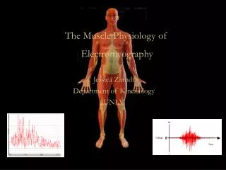
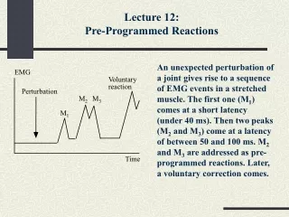
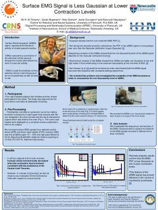
Surface EMG Signal is Less Gaussian at Lower Contraction Levels Ali H. Al-Timemy1, Guido Bugmann1, Nick Outram2, Javier Escudero2 and Kianoush Nazarpour3 1Centre for Robotics and Neural Systems, University of Plymouth, PL4 8AA, UK 2Signal Processing and Multimedia Communication (SPMC), University of Plymouth, UK 3Institute of Neuroscience, School of Medical Sciences, Newcastle University, UK E-mail: ali.ali@plymouth.ac.uk Institute of Neuroscience • Introduction • Surface Electromyogram (sEMG) signal represents the electrical activity of closely spaced muscles. • EMGs represents a summation of the electrical activity passing through the muscle and reflects the level of muscular activity. • EMG is used for prosthetic control, assistive device control because of its non invasiveness as well as ease of use. Background • Gaussian density function can model the EMG PDF [1]. • But during low intensity isometric contractions, the PDF of the sEMG signal is more peaked near zero than the Gaussian distribution (super-Gaussian) [2]. • Negentropy analysis of the EMGs showed that the non-Gaussianity level of the sEMG signal depends on the muscular contraction level [3]. • Bicoherence analysis of the EMGs showed that EMGs are highly non-Gaussian at low and high levels of force while being in the maximum Gaussianity at the mid-level of MVC [4]. • But Hussian et al. [5] used the bicoherence index and showed that the sEMG signal becomes less Gaussian with increased walking speed force. • We revisited this problem and investigated the suitability of the EMG bicoherence index to characterize the non-Gaussianity level of sEMG. Method 1. Participants. -Four right-handed subjects (two females and two males) participated in the study. The study was approved by the local ethics committee at Newcastle University. 2. Pre-Processing - Participants controlled a myoelectric cursor by making isometric contractions of right upper-limb muscles with the arm strapped to the chair armrest and the hand restrained in a glove which was fixed to the chair (Fig.1). The cursor and targets were displayed on a computer screen positioned in front of the subject. We recorded surface EMG signals from abductor policisbrevis (APB) and flexor carpi radialis(FCR) muscles. EMG was first amplified (gain 1K – 10K) and high-pass filtered at 30 Hz (Neurolog NL824/820, Digitimer) before sampling at 10 KHz (PCI-6071E, National Instruments). Fig. 1 Hand fixation for the experimental protocol Fig3. The subject performing 50% MVC with the visual feedback At the start of the experiment, subjects were informed of the structure of the task (Fig. 2) Control signals were computed by smoothing the rectified EMG to determine the cursor position along a 1D vertical axis. Visual feedback was provided using the computer monitor. We recorded the EMGs in six blocks each subject for each muscle in a range of the force levels 3. Data Analysis: We investigated the bispectrum and kurtosis of the EMG measurements to explore the features of the EMG signals recorded in different force level conditions. Fig2 . The myoelectric-controlled interface. • Conclusions • Kurtosis analysis results confirm that the EMG PDF is less Gaussian at low muscle contraction levels. • This feature of the sEMG signal has proved effective in the control of myoelectric prostheses [3]. • Results • In all four subjects and for both muscles, kurtosis values monotonically decreased when the contraction level increased consistent with the predictions of the Central Limit Theorem. • However, in contrast to [4] and [5], we did not observe any modulation of the bicoherence index with respect to muscle activity. Fig4 Kurtosis plot of FCR muscle for 4 subjects with standard deviation Fig5. Bispectrum plot for FCR muscle of 4 subjects with standard deviation References [1]E.A. Clancy and N. Hogan, “Probability density of the surfaceelectromyogram and its relation toamplitudedetectors,” IEEE Transactions onBiomedicalEngineering, vol. 46, (no. 6), pp. 730-739, 1999. [2]I. Hunter, R. Kearney, and L. Jones, “Estimation of the conductionvelocity of muscleactionpotentialsusingphase and impulseresponsefunctiontechniques,” Medical and BiologicalEngineering and Computing, vol. 25, (no. 2), pp. 121-126, 1987. [3]K. Nazarpour, A.R. Sharafat, and S.M.P.Firoozabadi, “Application of HigherOrderStatisticstoSurfaceElectromyogramSignalClassification,” IEEE Transactions onBiomedicalEngineering, vol. 54, (no. 10), pp. 1762-1769, 2007. [4]P.A. Kaplanis, C.S. Pattichis, L.J.Hadjileontiadis, and S.M. Panas, “Bispectral analysis of surface EMG,” in Mediterranean Electrotechnical Conference MELECON, 2000. [5]M.S. Hussain, M.B.I.Reaz, F. MohdYasin, and M.I. Ibrahimy, “Electromyographysignalanalysisusingwavelettransform and higherorderstatisticstodeterminemusclecontraction,” Expert Systems, vol. 26, (no. 1), pp. 35-48, 2009. Acknowledgments -This work is supported by the Iraqi Ministry of higher Education and Scientific Research / Cultural and scholarship Directorate Scholarship. - KN’s work is supported by The Leverhulme Trust and The Medical Research Council, UK.