Download
1 / 50
500 likes | 579 Views
Learn about renal blood flow regulation, glomerular function, GFR, and stages of renal insufficiency. Get insight into assessment approaches for patients with renal disease.

E N D
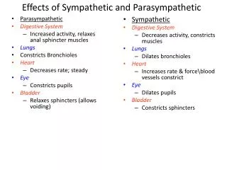
The afferents carrying sensations of stretch and fullness of the bladder are parasympathetic, • whereas pain,touch, and temperature sensations are carried by sympathetic nerves. • Sympathetic fibers are predominantly a-adrenergic in the bladder base and urethra, and ß-adrenergic in the bladder dome and lateral wall.
The kidneys receive approximately 15% to 25% of total cardiac output, or 1 to 1.25 L of blood per minute through the renal arteries, depending on the state of the body. Most of the blood is received by the renal cortex, with only 5% of cardiac output flowing through the renal medulla, which makes the renal papillae vulnerable to ischemic insults.
Renal blood flow is regulated by various mechanisms that control the activity of vascular smooth muscle and alter vascular resistance. • Sympathetic tone of renal vessels increases during exercise to shunt renal blood flow to exercising skeletal muscle; similarly, renal blood vessels relax during the resting condition of the body.
Sympathetic stimulation as a result of surgery can increase vascular resistance and reduce renal blood flow, whereas anesthetics may reduce renal blood flow by decreasing cardiac output.
Glomerular capillaries separate afferent arterioles from efferent arterioles. • Glomerular capillaries are highpressure systems, whereas peritubular capillaries are low-pressure systems. • Consequently, the glomerular capillaries are a fluid-filtering system, whereas the peritubular capillaries are a fluid-absorbing system.
vasa recta, a specialized portion of peritubular capillaries formed from efferent arterioles, are important in the formation of concentrated urine by a countercurrent mechanism. An intrinsic mechanism that causes • vasodilation and vasoconstriction of renal afferent arterioles regulates the autoregulation of renal blood flow.
A decrease in mean arterial pressure also decreases renal blood flow and eventually affects the glomerular filtration rate (GFR) when the pressure decreases to less than 60 mm Hg. • A persistently low mean arterial • pressure greater than 60 mm Hg affects renal blood flow, but does not affect the GFR because of the intrinsic mechanism of autoregulation
. Autoregulation maintains mean arterial pressure between 60 mm Hg and 160 mm Hg in intact and denervated kidneys.
Evaluation of Renal Function • Renal disease may be discovered incidentally during a routine medical evaluation, or patients may present with evidence of renal dysfunction, such as hypertension, edema, nausea, and hematuria • The initial approach in both situations should be to assess the cause and severity of renal abnormalities.
In all cases, this evaluation includes • (1) an estimation of disease duration, • (2) a careful urinalysis, and • (3) an assessment of the GFR.
Further diagnostic categorization is according to anatomic distribution: prerenal disease, postrenal disease, • and intrinsic renal disease.
Intrinsic renal disease can be divided further into • glomerular, • tubular, • interstitial, • vascular abnormalities. Laboratory tests useful in evaluating renal function
The GFR is by far tile best measure of glomerular function. • Normal GFR is around l25 mL/min. • However, manifestations of reduced GFR are not seen until the GFR has decreased to( 50%)of normal. • When GFR decreases to 30% of normal, a stage of moderate renal insufficiency sets in.
patients remain asymptomatic with only biochemical evidence of a decline in GFR (i.e., an increase in serum concentrations of urea and creatinine). • Further workup usually reveals other abnormalities, such as nocturia, anemia, loss of energy, decreasing appetite, and abnormalities in calcium and phosphorus metabolism.
As the GFR decreases further, a stage of severe renal insufficiency begins. • This stage is characterized by profound clinical manifestations of uremia and biochemical abnormalities, such as acidemia; volume overload; and neurologic, cardiac, and respiratory manifestations.
At the stages of mild and moderate renal insufficiency, intercurrent clinical stress may compromise renal function further and induce signs and symptoms of overt uremia.
When the GFR is 5% to 10% of normal, it is called end-stage renal disease (ESRD), and continued survival without renal replacement therapy becomes impossible. • Although most clinical abnormalities of corticotropin-releasing factor (CRF) are reversed by renal transplantation, the response to dialysis is quite variable
The blood urea nitrogen (BUN) concentration is not a direct correlate of reduced GFR. • BUN is influenced by nonrenal variables, such as exercise, bleeding, steroids, and massive tissue breakdown. • The more important factor is that BUN is not elevated in kidney disease until the GFR is reduced to almost 75% of normal.
Measurements of creatinine provide valuable information regarding general kidney function. • Creatinine in serum results from turnover of muscle tissue and depends on daily dietary intake of protein. • Normal values are 0.5 to 1.5 mg/100 mL; values of 0.5 to 1 mg/100 mL are present during pregnancy.
Creatinine is freely filtered at the glomerulus, and apart from an almost negligible increase in content because of secretion in the distal nephron, it is neither reabsorbed nor secreted.
Serum creatinine measurements reflect glomerular function and creatinine clearance is a specific measure of GFR.
This value should be multiplied by 0.85 for women because a lower fraction of body weight is composed of muscle.
The serum creatinine concentration and clearance are better indicators of general kidney function and GFR than similar measurements of urea nitrogen
Table 65-4 -- Conditions Affecting Blood Urea Nitrogen (BUN) Independently of Glomerular Filtration • Rate • Increased BUN • Reduced effective circulating blood volume (prerenal • azotemia) • Catabolic states (gastrointestinal bleeding, corticosteroid use) • High-protein diets • Tetracycline • Decreased BUN • Liver disease • Malnutrition • Sickle cell anemia • SIADH • SIADH, syndrome of inappropriate secretion of antidiuretic hormone.
Table 65-5 -- Conditions Affecting Serum Creatinine Independently of Glomerular Filtration Rate • Condition Mechanism • Conditions Causing Elevation • Ketoacidosis Noncreatinine chromogen • Cephalothin, cefoxitin Noncreatinine chromogen • Flucytosine Noncreatinine chromogen • Other drugs—aspirin, cimetidine, probenecid, • trimethoprim Inhibition of tubular creatinine secretion • Conditions Causing Decrease • Advanced age Physiologic decrease in muscle mass • Cachexia Pathologic decrease in muscle mass • Liver disease Decreased hepatic creatine synthesis and cachexia • Tubular Function
Important Pathophysiologic Manifestations ofChronic Renal Failure
Hypervolemia • Total-body contents of Na++ and H2O are increased in chronic renal failure (CRF), although this increase may not be clinically apparent until the GFR is reduced to very low levels. • Weight gain is usually associated with volume expansion and is offset by the concomitant loss of lean body mass.
Acidemia • Although urine can be acidified normally in most patients with CRF, these patients have a reduced ability to produce ammonia. • In the early stages, the accompanying organic anions are excreted in urine, and the • metabolic acidosis is of the non–anion gap variety.
With advanced renal failure, a fairly large “anion gap” may develop (to approximately 20 mmol/L), however, with a reciprocal decrease in plasma HCO3 concentration. • This acidemia is usually corrected by hemodialysis. • Although acidemia is well compensated in moderate CRF, patients can become acidemic and hyperkalemic in the postoperative period
Hyperkalemia • The approximate daily filtered load of K+ is 700 mmol. Most of this filtered load is reabsorbed in tubule segments, • most of the K+ excreted in the final urine reflects events governing K+ handling at the level of the cortical collecting tubule and beyond. • K+ excretion in the gastrointestinal tract is augmented in patients with CRF.
Hyperkalemia may be precipitated, however, in numerous clinical situations, including protein catabolism, hemolysis, hemorrhage, transfusion of stored red blood cells, metabolic acidosis, and exposure to various medications that inhibit K+ entry into cells or K+ secretion in the distal nephron.
Cardiac and Pulmonary Manifestations • Hypertension is a common complication of CRF and ESRD. • Because hypervolemia is the major cause of hypertension in uremia, normotension is usually restored by the use of diuretics in predialysis patients or by dialysis in ESRD patients.
Patients generally have left ventricular hypertrophy and accelerated atherosclerosis (disordered glucose and fat metabolism). • Pericarditis can be observed in underdialyzed patients versus patients with CRF who undergo regular dialysis.