Probabilistic Atlas of Thalamic Connectivity to Prefrontal Cortex
10 likes | 114 Views
Explore human thalamic architecture using diffusion MRI data to predict thalamic nuclei based on cortical parcellation. Understand thalamo-cortical circuitry disruptions. Utilize the atlas to assign functional-anatomical labels and investigate structure-function correlations.

Probabilistic Atlas of Thalamic Connectivity to Prefrontal Cortex
E N D
Presentation Transcript
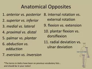
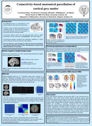
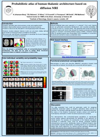
FMRIB C M1 PMC S1 PPC PFC OCC TEMP 1 0 A. Thalamus. B. Connectivity distribution from a seed voxel in MD nucleus terminates in the prefrontal cortex. C. Axial slice from histological atlas. D. Predicted classification of thalamic nuclei according to cortical parcellation shown in E. F. Classification of thalamic voxels according to the cortical region with which they have maximum probability of connection.. D A C B E F S1 M1 PMC PFC PPC TEMP OCC 100% 83% 66% 50% Probabilistic atlas of human thalamic architecture based on diffusion MRIH Johansen-Berg1, TEJ Behrens1, E Sillery1, O Ciccarelli2, A Thompson2, SM Smith1, PM Matthews11Oxford Centre for FMRI of the Brain, University of Oxford, UK2Institute of Neurology, Queen’s square, London, UK Methods. Diffusion-weighted data were acquired in 11 subjects2. From each thalamic voxel, we generated connectivity distributions3, and recorded probability of connection to each of 7 cortical masks. We classified seed voxels according to the cortical regions with which they showed the highest connection probability, resulting in exclusive connectivity-defined regions (CDRs). Individual subject CDRs (in standard space) were thresholded (>25% probability of connection), binarised and overlaid to create a probabilistic atlas. • Introduction • Thalamic activation seen during sensory, motor and cognitive tasks. • Thalamic lesions affect sensory, executive and memory functions . • Thalamo-cortical circuitry disrupted in neurological/psychiatric disorders. • However, borders between thalamic nuclei can only been reliably defined post mortem so structure-function correlations are limited. • We previously used diffusion tractography to parcellate the human thalamus in vivo based on its cortical connectivity1. • Here, we present a probabilistic atlas of connectivity-based thalamic architecture. The atlas can be used to assign likely functional-anatomical labels to thalamic activations or lesion sites. Web-based interface http://www.fmrib.ox.ac.uk/connect Inter-individual variability and probability maps Functional-anatomical correspondence Meta-analysis of motor and executive fMRI/PET activations showed close correspondence between functionally and connectivity-defined regions Top. Thalamic segmentation for 11 individual subjects Middle: Population probability maps showing extent of overlap across subjects. Maps were created by thresholding (at >25% of cortical pathways reaching that area) and binarising clusters for each cortical area and each subject. These binarised clusters were then overlaid so that the intensity in the group maps reflects the number of subjects showing connectivity from that voxel. Bottom:Axial slices through the whole thalamus showing edges of thresholded (at >4/11 subjects) group probability maps for connection to each cortical region. A B C Functional correspondence: Pale grey surface represents thalamus A. Thalamic activation sites during motor (blue) or executive (red) tasks. B. Co-localisation between motor activations and thalamic volumes with high probability of connection to motor (blue), premotor (red) or somatosensory (green) cortices. C. Co-localisation between executive activations and thalamic volumes with high probability of connection to prefrontal cortex. Connectivity profiles for activation centres. Each row represents one activation centre, each column represents connection to one cortical target. The brightness of each cell represents the probability of connection to that cortical target. Movement-related activations tend to have high probabilities of connection to sensorimotor, premotor and parietal cortices. Executive and memory tasks tend to have very specific high probability of connection to prefrontal cortex. Discussion The atlas presented here could be particularly useful for assigning probabilistic anatomical labels to thalamic activations when circuitry is unknown. For example, although thalamic activation is observed during pain, the nature of the thalamic involvement remains unclear and would be illuminated by clarification of which functional subunits are activated. The demonstration that connectivity-based parcellation is reproducible across healthy subjects demonstrates the feasibility of this approach for clinical studies. For example, the size/location of the thalamic sub-region connected to prefrontal cortex could be directly compared between schizophrenics and controls. Additionally, lesions of different thalamic sites cause specific cognitive impairments4. Localising lesions on our connectivity-based atlas would determine the thalamo-cortical pathways affected and enable more precise clinico-anatomical correlations to be made. Acknowledgements. UK MRC (PMM, SMS, ES), UK EPSRC (TEJB, SMS), Wellcome Trust (HJB), References 1. Behrens et al, 2003, Nature Neurosci 6, 750-7. 2. Wheeler-Kingshott et al, 2002, ISMRM 1118. 3. Behrens et al, 2003, MRM 50, 1077-88. 4. Van der Werf, 2003, Neuropsychologia, 41, 1330-1344.