Download
1 / 42
440 likes | 577 Views
Surgical Breast Pathology. Juan C. Cendan, MD Assistant Professor of Surgery. Objectives of Lecture. Categorize risk factors for cancer Describe diagnostic workup for breast masses and tools available to the clinician

E N D
Surgical Breast Pathology Juan C. Cendan, MD Assistant Professor of Surgery
Objectives of Lecture • Categorize risk factors for cancer • Describe diagnostic workup for breast masses and tools available to the clinician • Provide up-to-date guidelines in the screening and diagnosis of breast masses • Brief review of surgical options and implications in patients with breast cancer
Assessment of Risk/History • Four major risks (increase RR by 4x): • Family history • 1st degree relatives • Age at diagnosis, BRCA1/2 risk • Atypical hyperplasia on prior biopsies • Personal breast cancer history • LCIS
Assessment of Risk/History • Four Minor Risk Factors: 1-2x RR • Early menarche • Long interval from menarche to 1st child • Nulliparity • Ovarian or endometrial cancer • Estrogen therapy after menopause
Physical Exam • Be systematic • Inspection of breasts: sitting up, then recumbent • “Strip method” • Nipples • Lymph nodes
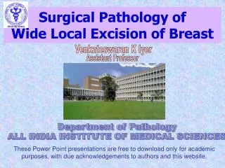
Diagnostics • Standard screening mammogram • CC and MLO • Diagnostic mammogram • Above, plus compression/additional views • In either case, 5-10% false negative and 90-95% sensitivity
Cranio-caudal (CC) view and mediolateral oblique (MLO) mammographic view
Diagnostics • Ultrasound • Useful in the young • Useful in pregnant women • Delineates solid vs cystic • MRI • Possibly the future of breast diagnostics, not there yet, limitations with biopsy
Biopsy techniques • Palpable solid mass • Needle or core biopsy • Incisional or excisional biopsy • Non-palpable mass • Stereotactic core • Stereotactic “mammotome” • Needle localized biopsy
Some Benign Conditions • Nipple Discharge • Incidence of malignancy when bloody (10-15%) and unilateral, though usually papilloma • More likely cystic or duct ectasia • Consider prolactin if bilateral
Benign, con’t • Fibroadenoma • Very common in young women • Freely mobile and smooth • Characteristic u/s appearance • Half of adenomas resolve if <3cm over 5yrs • Large adenomas should be biopsied to exclude rare phylloides tumor
Benign, con’t • Cysts • Due to relative excess estrogen, usually in 4-5th decades • Fluctuate with menses • Aspirate, if bloody then excise, send fluid for path the first time
Benign, con’t • Abscess, • Usually in lactating women • Painful and erythematous • Usually staph and strep • Drainage and antibiotics indicated • Rarely, can aspirate and treat with antibiotics • Caveats, in nonlactating (Ca), non-resolving (atypical infection), inflammatory cancer
Cancer • Most women with breast cancer have no risk factors! • Role of dietary fat, estrogen • Breast cancer genes responsible for 3-5% only
Cancer • DCIS • Carcinoma in situ • Usually found on mammography as microcalcifications • Felt to progress to invasive in 30-50% if untreated • Subtypes: comedo highest risk
Cancer • DCIS, con’t • Treatment • Non-invasive, so risk of LN disease is minimal • Must treat the breast, options: • Excise with large enough margins (>1cm) in a small tumor • Or, Excise and radiate • Or, Mastectomy +/- reconstruction
Cancer • Invasive Ductal Cancer • “Garden variety breast cancer” • More often presents with mass than DCIS • Treatment: • BREAST: Excise and RT or mastectomy, Cannot just excise with margins (30-40% recur) • Lymph Nodes: Must be sampled for staging • Sentinel Node vs Axillary Dissection
Cancer • Chemotherapy • Recommended for tumors >1cm in most patients • Recommended if lymph nodes are positive • 8 recommended chemo protocols at this time!! • ER positivity and Tamoxifen
Cancer • Survival