Cardiovascular System
570 likes | 688 Views
Learn about the characteristics of blood, blood volumes, cell types, hemoglobin, white blood cells, platelets, plasma, and more in the cardiovascular system. Understand how blood transports substances, its volume variances, and the importance of different blood cell types.

Cardiovascular System
E N D
Presentation Transcript
Characteristics of Blood • Connective tissue • Plasma and cells • Transports substances between body cells and the external environment
Blood Volumes • Varies with body size, fluid and electrolyte concentrations, and amount of adipose • Average adult volume – 5 liters (4-5 L for women, 5-6 L for men) • Hematocrit – usually 45%
Blood Cell Types • Erythrocytes – RBCs • Leukocytes – WBCs • Thrombocytes - platelets
Red Blood Cells (RBCs) • Biconcave disks that thin near the centers increased surface area for transporting gases • Have nuclei early in development, but extrude them • No nucleus – more room for hemoglobin, but cannot reproduce or make proteins
Hemoglobin • Oxygen-carrying protein • 1/3 of each RBC • Gives blood its color • Oxyhemoglobin • Deoxyhemoglobin • Hypoxia • Cyanosis
Red Blood Cell Counts • RBCC – the number of RBCs in 1 mm3 of blood • Adult male average – 4.6 – 6.2 million • Adult female average – 4.2 – 5.4 million • Determines blood’s oxygen carrying capacity • Important diagnostic tool
Blood Cell Production • RBCs are normally flexible, elastic, and able to pass through small blood vessels • More fragile as they age • Macrophages phagocytize and destroy damaged RBCs in the liver and spleen
White Blood Cells (WBCs) • Function to protect against disease • Phagocytize bacterial cells • Produce antibodies • Move by diapedesis • 2 main types: • Granulocytes – neutrophils, eosinophils, and basophils • Agranulocytes – monocytes and lymphocytes
Granulocytes • Lobed nucleus with 2-5 sections • Dark staining nucleus and pale granules • 54-62% of WBCs • Contain many lysosomes – actively phagocytizes bacteria
Granulocytes • Contains coarse, uniformly sized granules • 2 lobes on nucleus • Stains red • 1-3% of WBCs • Kills parasites • Helps control inflammation and allergic reactions
Granulocyte • Fewer, more irregular granules than eosinophils • Granules stain deep blue • Less than 1% of WBCs • Contain heparin (inhibits blood clotting) and histamine (increases blood flow to injured tissues)
Agranulocytes • Largest blood cells • May live weeks months • 3-9% of WBCs • Discussed later
Agranulocyte • Form in red bone marrow and lymphatic system • Slightly larger than RBCs • Large, round nucleus with rim of cytoplasm • May live for years • 25-33% of WBCs • Discussed more later
White Blood Cell Count (WBCC) • Number of WBCs in 1 mm3 of blood • Adult average – 5000 – 10,000 cells • Leukocytosis • Leukopenia • Differential WBCC (DIFF) • More neutrophils – bacterial infection • More eosinophils – parasitic infection, allergic reaction • Leukemia
Platelets • Not complete cells • Form from megakaryocytes that fragment • Lack nuclei • Half size of RBCs • Live 10 days • Average count – 130,000 – 360,000 cells • Help close breaks in damaged blood vessels • Initiate formation of blood clots
Plasma • 92% water • Functions include: • Transporting nutrients, gases, and vitamins • Regulating fluid and electrolyte balance • Maintaining pH
Hemostasis • Vasospasm • Platelet plug formation • Coagulation • Fibroblasts invade clot
Blood Groups • ABO blood group is based on the presence or absence of 2 major antigens (RBC surface molecules) • 4 possible combinations: • A • B • AB • O
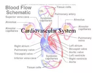
System Overview • System consists of the heart and the blood vessels • Function: to bring oxygen and nutrients to all body cells and to remove waste
Coverings of the Heart • Pericardium • Fibrous pericardium • Parietal pericardium • Visceral pericardium • Pericardial cavity • Pericardial fluid reduces friction
Walls of the Heart • Epicardium • Myocardium • Endocardium • Purkinje fibers
Heart Chambers • 4 chambers of the heart • Atria • Thin walls • Receive blood returning to the heart • Auricles • Ventricles • Thicker walls • Receive blood from atria • Force blood out of heart • Septum
Heart Valves • Atrioventricular valves • Tricuspid – right • Bicuspid – left • Semilunar valves • Pulmonary – right • Aortic – left • Chordae tendinae • Papillary muscles
Blood Supply to the Heart • Coronary arteries • Cardiac veins • Coronary sinus
Cardiac Cycle • Cardiac cycle – series of events that constitute a complete heartbeat • Systole – contraction • Diastole – relaxation
Cardiac Cycle • Heart Sounds – “lub-dup” • Lub – ventricular systole • AV valves close • Dup – ventricular diastole • SL valves close • Murmur
Cardiac Conduction System • Coordinates the events of the cardiac cycle • Consists of clumps and strands of specialized cardiac muscle that initiate and distribute impulses throughout the myocardium
Nodes of Cardiac Conduction System • Sinoatrial node – AKA “pacemaker” • Just beneath epicardium • Located in right atrium near opening of superior vena cava • Initiates impulses without nervous stimulation
Nodes of Cardiac Conduction System • Atrioventricular node • Located in inferior portion of septum • AV bundle (bundle of His) • Large fibers run through the interventricular septum • Purkinje fibers • Spread from septum into papillary muscles • Stimulate ventricular contraction
Regulation of Cardiac Cycle • Parasympathetic and sympathetic fibers from medulla oblongata run to the nodes • Secrete acetylcholine to decrease heart rate • Secrete norepinephrine to increase heart rate • Cardiac center
Electrocardiogram (ECG) • Recording of the electrical changes in the myocardium during the cardiac cycle • P – atrial systole • QRS – ventricular systole; covers atrial diastole • T – ventricular diastole
Arteries • Strong, elastic vessels that carry blood away from the heart • Lead to finer branches called arterioles


