Epilepsy
Epilepsy. Dr. Hawar A. Mykhan M.B.Ch.B ., F.I.B.M.S. Seizures. A seizure is a paroxysmal event due to abnormal, excessive, hypersynchronous discharges from an aggregate of CNS neurons.

Epilepsy
E N D
Presentation Transcript
Epilepsy Dr. Hawar A. Mykhan M.B.Ch.B., F.I.B.M.S
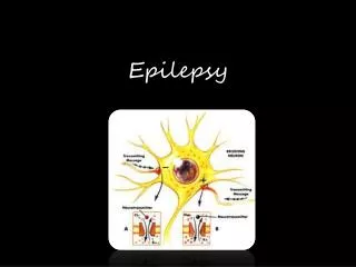
Seizures • A seizure is a paroxysmal event due to abnormal, excessive, hypersynchronous discharges from an aggregate of CNS neurons. • Epilepsy is diagnosed when there are two or more unprovoked seizures due to a chronic, underlying process. • The first step in the evaluation is to determine whether the seizure was provoked or unprovoked. • Provoked seizures are caused by factors that can disrupt cerebral function.
Possible precipitating factors: • Drug ingestion or withdrawal. • Structural lesions of the brain. • Physical injuries. • Vascular insults. • Infections. • metabolic or toxic abnormalities. • Acute symptomatic seizure: happens within 1 week of the precipitating factor. • Remote symptomatic seizure: More than 1 week of the precipitating factor.
A seizure that occurs in the absence of any identifiable factor is an unprovoked seizure. • Idiopathic seizures: There is no identifiable cause. • Cryptogenic seizure: Suspected to be symptomatic, but no symptomatic cause identified on imaging. • If the seizure is caused by a specific provoking factor and that factor can be corrected, no further treatment is required. • Management of unprovoked seizures is more complicated, and the risks of recurrent seizure need to be assessed and discussed with the patient and family.
The second step is to determine whether the seizure was generalized or focal. • Focal seizures are associated with a focal brain lesion and require a thorough investigation with imaging and are also a risk factor for recurrent seizure activity. • Studies have suggested that the risk of a recurrent seizure after the first unprovoked seizure is 30% to 50%. • After a second unprovoked seizure, this risk increases to 70% to 80% and treatment with antiepileptic drugs is often initiated at this time.
Seizure Classification • This is essential for diagnosis, therapy, and prognosis. • Seizures are partial or generalized. • partial seizures originate in a localized area of cortex and generalized seizures involve diffuse regions of the brain in a bilaterally symmetric fashion. • Simple-partial seizures do not affect consciousness and may have motor, sensory, autonomic, or psychic symptoms. • Complex-partial seizures include alteration in consciousness coupled with automatisms (e.g., lip smacking, chewing, aimless walking, or other complex motor activities).
Generalized seizures may occur as a primary disorder or result from secondary generalization of a partial seizure. • Tonic-clonicseizures (grand mal) cause sudden loss of consciousness, loss of postural control, tonic muscular contraction producing teeth-clenching and rigidity in extension (tonic phase), followed by rhythmic muscular jerking (clonic phase). Tongue-biting and incontinence may occur during the seizure. • Recovery of consciousness is typically gradual over many minutes to hours. Headache and confusion are common postictal phenomena.
Etiology • Neonate (<1 month): Hypoxia, CNS infection, metabolic, drug withdrawal, developmental and genetic disorders. • Infants and children (<12 years): Febrile, genetic, CNS infection, trauma and idiopathic. • Adolescent and young adults (13-35): Trauma, alcohol withdrawal, drugs, tumor, idiopathic. • Older adults (>35):Cerebrovascular disease (50%), tumor, alcohol withdrawal, metabolic, Idiopathic.
Clinical Evaluation • Careful history is essential since diagnosis of seizures and epilepsy is often based solely on clinical grounds. • Differential diagnosis includes syncope or psychogenic seizures (pseudoseizures). • General exam includes search for infection, trauma, toxins, systemic illness, neurocutaneous abnormalities, vascular disease and drugs. • Asymmetries in neurologic exam suggest brain tumor, stroke, trauma, or other focal lesions.
Laboratory Evaluation • CBC, electrolytes, serum glucose, liver and renal function, urinalysis, and toxicology screen. • A lumbar puncture is indicated if there is any suspicion of CNS infection such as meningitis or encephalitis. This is mandatory in HIV-infected patients even in the absence of symptoms or signs suggesting infection.
Electroencephalography (EEG) • All patients should be evaluated as soon as possible with an EEG, which measures electrical activity of the brain by recording from electrodes placed on the scalp. • The presence of electrographic seizure activity during the clinically evident event establishes the diagnosis. • The absence of electrographic seizure activity does not exclude a seizure disorder. • The EEG can show abnormal discharges during the interictal period that support the diagnosis of epilepsy, and is useful for classifying seizure disorders and determining prognosis.
Brain Imaging • All patients with unexplained new-onset seizures should have a brain imaging study (MRI) to search for an underlying structural abnormality. • The only exception may be children who have an unambiguous history and examination suggestive of a benign, generalized seizure disorder such as absence epilepsy.
Treatment • Acutely, the patient should be placed in semiprone position with head to the side to avoid aspiration. • Tongue blades or other objects should not be forced between clenched teeth. • Oxygen should be given via face mask. • Reversible metabolic disorders (e.g., hypoglycemia, hyponatremia, hypocalcemia, drug or alcohol withdrawal) should be promptly corrected. • Longer-term therapy includes treatment of underlying conditions, avoidance of precipitating factors, prophylactic therapy with antiepileptic medications or surgery, and addressing various psychological and social issues.
Choice of antiepileptic drug therapy depends on a variety of factors including seizure type, dosing schedule, and potential side effects. • Therapeutic goal is complete cessation of seizures without side effects using a single drug (monotherapy) and a dosing schedule that is easy for the patient to follow. • If ineffective, medication should be increased to maximal tolerated dose based primarily on clinical response rather than serum levels.
If unsuccessful, a second drug should be added, and when control is obtained, the first drug can be slowly tapered. • Some patients will require polytherapy with two or more drugs, although monotherapy should be the goal. • Patients with certain epilepsy syndromes (e.g., temporal lobe epilepsy) are often refractory to medical therapy and benefit from surgical excision of the seizure focus.
Generalized Tonic-Clonic (Grand Mal) Epilepsy • Accounts for 10% of all epilepsy and is the most common metabolic seizure. • Tonic phase: lasts 10-20 seconds and is often started by “ictal cry,” due to contraction of expiratory muscles. It causes cyanosis, tongue biting, increased HR, BP and pupil size. • Clonic phase: intermittent relaxation periods produced by the superimposition of periods of muscle relaxation on the tonic muscle contraction which get progressively longer ( about one minute). • Postictal: patients become unresponsive, flaccid, with excessive salivation, headache, fatigue, bladder or bowel incontinence.
Absence (Petit Mal) Epilepsy • Absence seizures are characterized by sudden, brief lapses of consciousness without loss of postural control. • The seizure typically lasts for only seconds, consciousness returns as suddenly as it was lost, and there is no postictal confusion. • Absence seizures usually begin in childhood (4–8 years) or early adolescence. • The seizures can occur hundreds of times per day. • EEG: 3 Hz spike and wave • Hyperventilation tends to provoke these electrographic discharges and even the seizures themselves and is routinely used when recording the EEG.
Benign Childhood Epilepsy with Centrotemporal Spikes (Rolandic, Sylvian Epilepsy) • Most common epilepsy syndrome of childhood. • Begins at age 5-9. • The seizures have a characteristic semiology, involving hemifacialclonic movements, speech arrest, dysarthria, and excessive drooling. • Remits by the age of 20. • EEG: spikes in the rolandic (motor strip) or centrotemporal area.
Benign Childhood Epilepsy with Centrotemporal Spikes (Rolandic, Sylvian Epilepsy)
Juvenile Myoclonic Epilepsy • The age of onset is 8-24(peak, in teens). • The patient is developmentally normal. • Seizure happens predominantly on awakening (early morning or nap). • Aggravated by sleep deprivation and photic stimulation. • Consciousness is preserved unless the myoclonus is especially severe. • Maybe associated with other seizure types like absence or grand mal epilepsy.
Mesial Temporal Lobe EpilepsySyndrome • It is the most common syndrome associated with complex partial seizures. • There is history of febrile seizures in childhood with positive family history of epilepsy. • Clinically aura is common with Behavioral arrest/stare, complex automatisms, postictal disorientation and memory loss. • High-resolution MRI can detect the characteristic hippocampal sclerosis. • It tends to be refractory to treatment with anticonvulsants but responds extremely well to surgical intervention.
Mesial temporal lobe is shrunken and malformed with enlargement of the temporal horn of the lateral ventricle.