Download
1 / 57
570 likes | 688 Views
This case presents a 54-year-old female with a 10-year history of chronic diarrhea exacerbated over the last year. Key symptoms involve severe episodes of diarrhea, abdominal pain, and weight loss. Investigations revealed thickened small intestine mucosa and matted loops. Despite trials of treatment including colestyramine and indomethacin withdrawal, no significant improvement ensued. The patient's history of endometrial carcinoma treated with radiotherapy raises suspicion of late-onset radiation enteritis, complicating her gastrointestinal symptoms.

E N D
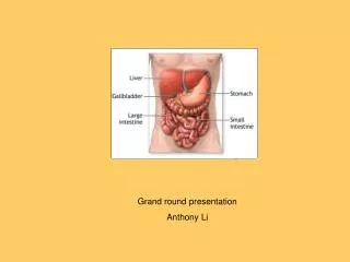
Grand round presentation Anthony Li
Mrs J D – 54 yrs ♀ • PC: • diarrhoea • HPC: • bowels ‘not right’ for 10 yrs • worse last 1 yr • BO normally: • x3 - 4 per day • firmish • floaty • some difficulty flushing • no associated abdominal pain / PR bleeding
Mrs J D – 54 yrs ♀ • HPC: • last 6 mths - x6 episodes of severe diarrhoea: • BO x9 in 24 hrs • associated with: • diffuse abdominal pain • vomiting x4 - 5 → unable to keep any PO intake down • no back pain / jaundice / change of colour of urine or stool • symptoms settle next day → feels ‘exhausted’ • no obvious precipitants • admitted to Crawley for 48 hrs with latest attack – no Ix performed • weight loss of approx. 1 st
Mrs J D – 54 yrs ♀ • PMH: • sterilisation • retained placenta • tonsillectomy • Hysterectomy(endometrial ca) • DH: • immodium 2 tabs tds • metoclopramide 1 tab tds • temazepam 40mg nocte • norval 30mg nocte • indomethacin 25mg tds
Mrs J D – 54 yrs ♀ • allergies: • NKDA • FH: • ? • SH: • occupation - home helper • smoker - 10/day • no EtOH • x3 children at home 18yrs, 15yrs, 12yrs
Mrs J D – 54 yrs ♀ • O/E: • General: • thin • no jaundice / anaemia / clubbing / lymphadenopathy • RS: • NAD • CVS: • NAD • Breasts: • NAD
Mrs J D – 54 yrs ♀ • O/E: • GI: non-distended visible SB segmentation centrally tender RUQ over GB - no guarding no palpable masses BS normal DRE: tender left lateral pelvic wall but NAD pale steatorrhoeic stool
Initial investigations • sigmoidoscopy: • 2 - 3 small telangiectases between 12 - 15 cms, otherwise normal to 15cms • bloods: • FBC, U&Es, LFTs, Ca2+, glu – WNL • TFTs, B12, folate – WNL • Inflammotory markers- WNL • Coeliac screen - negative • stool: • 3 day faecal fats – marginally ↑ at 11 g/day ( up to 7.5 g/day ) • swab – no salmonella, shigella or campylobacter • USS abdo: • NAD – no gallstones
Further investigations • Therapeutic trial with colestyramine did not help • Indomethacin withdrawal did not work • Test for SBBO was negative • Faecal elastase was normal • SBFT showed-
transverse barring from thickened valvulae conniventes- stack of coin appearance
IT’S ALL ABOUT THIS! DEB GHOSH GASTRO SPR
Any Guess? A 54 yr old lady presents with chronic diarrhoea with thickened SI mucosa, stricture and matted loops
Further history • Endometrial carcinoma treated with post-op radiotherapy 10years back- weighed 6 stone at time of radiotherapy • Severe diarrhoea two weeks post radiotherapy lasting for couple of weeks • Mild symptoms only for next ten years
OVERVIEW OF MANAGEMENT OF DIARRHOEA FOR NON -GASTROENTEROLOGIST
What is diarrhoea? • Abnormal passage of 3 or more loose or liquid stools per day for > 4weeks and / or a daily stool weight greater than 200g/day
Major causes • Irritable bowel syndrome • Inflammatory bowel disease • Chronic infections • Malabsorption syndromes Typical symptoms, normal exam and normal screening blood tests- no further investigations needed
Major causes • Irritable bowel syndrome • Inflammatory bowel disease • Chronic infections • Malabsorption syndromes
Major causes Irritable bowel syndrome Inflammatory bowel disease Chronic infections Malabsorption syndromes
Minor causes • Ischaemic colitis • Drugs • Neoplastic • Motility disorders • Radiation enteritis Incidence of ischemic colitis at various locations (%) • Descending colon 37 • Splenic flexure 33 • Sigmoid colon 24 • Transverse colon 9 • Ascending colon 7 • Rectum 3
Minor causes Ischaemic colitis Drugs Neoplastic Motility disorders Radiation enteritis
Minor causes Lymphoma Villous adenoma Gastrinoma VIPoma carcinoid • Ischaemic colitis • Drugs • Neoplastic • Motility disorders • Radiation enteritis
Minor causes • Ischaemic colitis • Drugs • Neoplastic • Motility disorders • Radiation enteritis Post surgical states- vagotomy/gastrectomy Endocrine- DM/Hyperthyroidism/carcinoid Infiltrative SI disease- scleroderma OCTT- Ba studies Radionucleotide scintigraphy
Minor causes • Ischaemic colitis • Drugs • Neoplastic • Motility disorders • Radiation enteritis Radiation of more than 50Gy Ileum and rectum mostly Mucosal damage and SBBO
Understanding of patient’s complain of diarrhoea • consistency • frequency of stools • urgency or faecal soiling • Stool characteristics • presence of visible blood- IBD or cancer • greasy stools that float and are malodorous -fat malabsorption
Duration of symptoms, nature of onset (sudden or gradual) • The volume of the diarrhoea • voluminous watery diarrhoea -small bowel • small-volume frequent diarrhoea -colon • Occurrence of diarrhoea during fasting or at night- secretory or organic diarrhoea
Travel history • Risk factors for HIV infection • Family history of IBD • Weight loss • Systemic symptoms as fevers, joint pains, mouth ulcers, eye redness-IBD • Previous therapeutic interventions- surgery and radiotherapy
A relevant dietary (sugar free products containing sorbitol and use of alcohol) • All medications (including over-the-counter drugs and supplements) • Association of symptoms with specific food ingestion (such as dairy products or potential food allergens) • A sexual history • anal intercourse-infectious proctitis • promiscuous sexual activity -HIV infection
Physical examination rarely provides a specific diagnosis. • Findings suggestive of IBD (eg, mouth ulcers, a skin rash, episcleritis, an anal fissure or fistula, the presence of visible or occult blood on digital examination, • Abdominal masses or abdominal pain, • Evidence of malabsorption (such as wasting, physical signs of anemia, scars indicating prior abdominal surgery), • Lymphadenopathy (possibly suggesting HIV infection), and • Abnormal anal sphincter pressure or reflexes (possibly suggesting fecal incontinence). • Palpation of the thyroid and examination for exopthalmus and lid retraction may provide support for a diagnosis of hyperthyroidism.
Basic laboratory evaluation • FBC • Thyroid function tests • ESR/CRP • U/E • Total protein and albumin, and • Ferritin/ folate/B12/Ca • Stool culture and microscopy
Treatment • General measures: • Hydration and electrolyte balance • Vitamins supplements • Loperamide (also improves bile acid absorption ) • Therapeutic trials • Colestyramine for BAM • Lactose free diet • Antibiotics for SBBO • For bleeding from proctitis in RE • Stool softener • Argon plasma coagulation • Formalin irrigation ( experimental )
RADIATION ENTEROCOLITIS Dr.E.M.Phillips
Historical aspects Self exposure Deep tissue traumatisation from Roentgen ray exposure Walsh,D: Br Med J 1897: 272 – 273 Animal experiments Roentgen ray intoxication. Warren S, Whipple GH: J Exp Med 1922: 35: 187 – 202 Post radiotherapy pathology38 patients Warren S, Friedman NB: Pathology and pathological diagnosis of radiation lesions in the gastrointestinal tract: Am J Path 1942: 499 – 513 1950s super voltage therapy 100 patients DeCosse JJ et al. Natural history & management of radiation induced injury of the gastrointestinal tract Ann Surg 1969; 170: 369 - 384
Symptoms Early During therapy and up to six months Late Five to 31 years after radiotherapy Peak onset 12 – 15 years after
Symptoms Diarrhoea Colic Nausea Mucosal Pathology Decrease: enterocyte turnover & villous height Increase: enterocyte death; mucosal oedema & inflammatory infiltrate with mucosal slough Early
Inflamm infiltrate and oedema Withering of crypts Cystic dilatation of crypt
Symptoms SB Diarrhoea/malabsorp’n Blind loop syndrome Subacute obstruction Colon tenesmus & mucus Bothhaemorrhage, fistula perforation Pathology Arteriolar endothelial spasm, damage & obliterative vasculitis Submucosa to serosa ischaemia, ulceration, and perforation; increase in bizarre fibroblasts; stricture, webs and fistula Late
Chronic Radiation Proctitis Vascular ectasia Thickening of lamina propria with fibrosis