Download
1 / 53
530 likes | 659 Views
The Reproductive System Chapter 26. Joe Pistack MS/ED. Functions. Reproductive system performs two functions: Produces, nurtures, and transports ova and sperm. Secretes hormones. Primary reproductive organs are the gonads. Female gonads---ovaries Male gonads------testes.

E N D
The Reproductive SystemChapter 26 Joe Pistack MS/ED
Functions • Reproductive system performs two functions: • Produces, nurtures, and transports ova and sperm. • Secretes hormones. • Primary reproductive organs are the gonads. Female gonads---ovaries Male gonads------testes
Male Reproductive System • Performs three functions: 1. Produces, nourishes, and transports sperm. 2. Deposits sperm within the female reproductive tract. 3. Secretes hormones.
Male Reproductive System • Testes-male gonads. • Functions: produce sperm and secrete the male hormone, testosterone. • Two oval testes are located outside the abdominal cavity and are suspended in a sac between the thighs called the scrotum. • Testes normally descend into the scrotum during the last 2 months of fetal development. • Cryptorchidism-failure of the testes to descend into the scrotum, can result in sterility.
Male Reproductive System • Undescended testicles are associated with infertility. • Sperm cannot live at body temperature, they prefer the cooler temperature of the scrotum. • Wearing tight underwear and jeans can elevate the temperature of the testes, thereby lowering sperm count.
Male Reproductive System • The testis is divided into about 250 smaller units called lobules. • Each lobule contains seminiferous tubules and interstitial cells. • Seminiferous tubules-tightly coiled tubules where sperm is produced. • The interstitial cells lie between the seminiferous tubules and produce the male hormones called androgens. • Most important androgen is testosterone. Testes produce sperm and testosterone.
Male Reproductive System • Spermatogenesis-the formation of sperm. • Each spermatogonium (undifferentiated sperm cell) contain 46 chromosomes, normal number of chromosomes for human body cells. • Under the influence of testosterone spermatogonium enlarge and form primary spermatocytes • Primary spermatocytes divide by a special type of cell division called meiosis. • Meiosis reduces the number of chromosomes by one half, a sperm will only have 23 chromosomes.
Male Reproductive System • When the sperm unites with an egg, which also has 23 chromosomes, the fertilized egg will have 46 chromosomes. • Newly formed sperm are not functional and must undergo several maturational changes.
Male Reproductive System • Sperm looks like a tadpole. • Sperm has: 1. a head 2. a body 3. a tail • Head is primarily a nucleus, contains the genetic information.
Male Reproductive System • Front part of the head contains enzymes that help the sperm penetrate the egg at the time of fertilization. • Body or midpiece is a spiral-shaped structure that contains mitochondria and supplies the sperm with energy for the “Big swim”.
Male Reproductive System • Tail of the sperm is the flagellum, has whip-like movements that enable the sperm to swim. • Most sperm live only hours after being deposited in the female reproductive tract, but hardier ones may live up to 3 days. • Purpose of the reproductive system is to produce offspring, this is achieved by the union of the sperm and the egg.
Male Reproductive System • As the sperm form, they gather in the seminiferous tubules and then move into a series of genital ducts, where they mature. • They are transported from the testes to the outside of the body. The ducts include: 2 epididymides 2 vas deferens 2 ejaculatory ducts 1 urethra
Male Reproductive System • Accessory glands-add secretions to the sperm as they travel through the genital ducts. • The three accessory glands are: 1. The seminal vesicles 2. The prostate gland 3. The bulbourethral glands
Male Reproductive System Prostate gland-single gland, donut-like, encircles the upper urethra just below the bladder. • Secretes a milky, alkaline substance that plays a role in increasing sperm motility. • Counteracts the acidic environment of the vagina and helps protect the sperm as it enters the woman’s reproductive tract.
Male Reproductive System • Semen-mixture of sperm and the secretions of the accessory glands. • About 60% comes from the seminal vesicles, the remainder comes from the prostate gland. • Semen is a milky white liquid with an alkaline pH. • Number of sperm per ejaculation is about 50 to 100 million.
Male Reproductive System • External genitals-consist of the scrotum and the penis. • Scrotum is the sac of skin that hangs loosely and contains the testes. • The penis has two functions: 1. Carries urine through he urethra to the outside of the body. 2. Acts as the organ of sexual intercourse.
Male Reproductive System • The loose skin covering the penis that extends downward and forms a cuff is called the foreskin or prepuce. • Circumcision is removal of the foreskin. • Phimosis-condition where the foreskin becomes tight and must be surgically removed.
Male Reproductive System • Male sex hormones are called androgens. • Primary male sex hormone is testosterone. • Most testosterone is secreted by the interstitial cells of the testes, small amount by the adrenal cortex. • Secretion begins during fetal development and continues at a low level throughout childhood.
Male Reproductive System • Puberty (age 10-13), testosterone secretion increases rapidly, transforming the boy into a man. • Testosterone is secreted continuously throughout life, and is responsible for the development of the male sex characteristics. • Primary sex characteristics include enlargement and development of the testis and various accessory organs such as the penis.
Male Reproductive System • Secondary sex characteristics refer to special features of the male body, such as: • Increased growth of hair on the face, chest, axillary and pubic region. • Deepening of the voice due to enlargement of the vocal cords. • Thickening of the skin and increased activity of the oil and sweat glands. • Increased musculoskeletal growth and development, broad shoulders and narrow waist.
Male Reproductive System • Hormones that control male reproductive system: • Primarily secreted by the hypothalamus, anterior pituitary gland, and the testes. • Hypothalamus secretes a releasing hormone • This stimulates the anterior pituitary gland to secrete: 1. follicle-stimulating hormone (FSH)-promotes spermatogenesis 2. luteinizing hormone (LH)-promotes development of interstitial cells and the secretion of testosterone
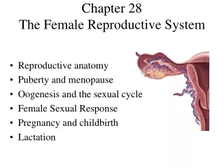
Female Reproductive System Functions: • Produces eggs • Secretes hormones • Nurtures and protects a developing baby during 9 months of pregnancy
Ovaries • Female gonads • 2 almond-shaped ovaries located on either side of the uterus in the pelvic cavity • Anchored in place by ligaments
Egg Development • Production of eggs begins at puberty and continues until menopause. • Supply of eggs exceeds actual need. • Each ovarian follicle consists of an immature egg. • Beginning at puberty, several follicles mature every month, but only one fully matures.
Egg Development • Within the ovary are saclike structures called ovarian follicles • Born with about 2 million follicles • By puberty only about 400,000 remain • Only 400 follicles ever fully mature • Typically one egg per month matures between puberty and menopause which occurs between 45 and 55 • Each ovarian follicle consists of an immature egg called an oocyte • As the egg matures it undergoes meiotic cell division which cuts the chromosomes from 46 to 23
Egg Development • As the follicle enlarges, a fluid-filled center is formed, and the follicular cells begin to secrete estrogen. • The mature ovarian follicle is known as the graafian follicle.
Ovulation • Once a month the ovarian follicle bursts. • The ovary ejects a mature egg (ovum) with a surrounding layer of cells. • The ejection phase is called ovulation.
Ovulation • The egg travels from the surface of the ovary into the peritoneal cavity and is immediately swept into the fallopian tubes. • Fimbriae-fingerlike projections at the end of the fallopian tubes that sweep the egg into the fallopian tubes.
Ovulation • If the egg is fertilized, it implants itself in the uterine lining and grows into a baby. • If the egg is not fertilized, it dies and is eliminated in the menstrual blood. • Once ovulation occurs, the follicular cells that remain in the ovary develop into a glandular structure called the corpus luteum (“yellow body”).
Ovulation • Corpus luteum secretes: 1. large amounts of progesterone 2. small amounts of estrogen • If fertilization does not occur, the corpus luteum deteriorates in about 10 days and becomes known as the corpus albicans (“white body”). • The dead corpus is no longer capable of secreting hormones.
Ovarian Cyst • Ovarian cysts occur when the corpus luteum fills with fluid. • A chocolate cyst occurs when the cyst is filled with blood. • Cysts may resolve on their own or they may require surgery.
Ovarian Hormones At puberty the ovaries begin to secrete estrogen and progesterone. Estrogen- • Promotes the maturation of the egg • Helps to develop the secondary sex characteristics • Gives the female the femininizing effects
Ovarian Hormones Feminizing effects of estrogen: • enlargement and development of the organs of the reproductive system. • Enlargement and development of the breasts. • Deposition of fat beneath the skin, especially in the thighs, buttocks, and breasts. • Widening of the pelvis. • Onset of the menstrual cycle. • Closure of the epiphyseal discs in the long bones.
Ovarian Hormones Progesterone- • works with estrogen in establishing the menstrual cycle. • Helps maintain pregnancy. • Prepares the breasts for milk production after pregnancy.
Genital Tract Consists of: • Fallopian tubes • Uterus • Vagina Fallopian tubes-also called uterine tubes or oviducts. • Each fallopian tube is about 4 inches long • Extend from either side of the uterus to the ovaries
Fallopian Tubes • Infundibulum-funnel-shaped end of the fallopian tube nearest to the ovary. • Fimbriae-fingerlike projections at the end of the fallopian tubes. • Fallopian tubes do not attach directly to the ovary, the fimbriae hang over the ovary.
Fallopian Tubes Functions: • Tube transports the egg from the ovary to the uterus. • The tube is the usual site of fertilization of the egg by the sperm.
Tube Troubles • Ectopic pregnancy-the fertilized egg implants in the fallopian tube rather than in the uterus. • Usually results in miscarriage, causes bleeding, possible hemorrhage, and sometimes death.
Tube Troubles Scarring of the fallopian Tubes : • Can be caused by repeated gonorrheal infections. • Blocks movement of the egg through the tube. • May cause sterility.
Pelvic Inflammatory Disease • Fallopian tubes open directly into the pelvic cavity. • Infection spreads through the tubes into the pelvic cavity, causing pelvic inflammatory disease (PID). • PID is most frequently associated with sexually transmitted diseases.
Uterus • Uterus or womb-shaped like an upside-down pear and is located between the urinary bladder and the rectum. • Broad ligament-holds the uterus in place.
Uterus Functions: • Provides a safe and nurturing environment for the growing baby. • Baby’s cradle for 9 months. • During pregnancy, the size of the uterus increases to hold the growing baby and the placenta.
Uterus Parts of the uterus: • Fundus-upper dome-shaped region above the entrance to the fallopian tubes. • Body-central region. • Crevex-lower narrow region that opens into the vagina.
Uterus Three layers: • Epimetrium-outer serosal layer. • Myometrium-middle, smooth, muscular layer. • Endometrium-inner layer, composed of two 2 layers,
Endometrium Inner layer, has 2 layers: 1. Basilar layer-thin and vascular, lies next to the myometrium. 2. Functional layer-responds to ovarian hormones, thickens in preparation for the fertilized egg. Layer that sloughs during menstruation. Site of Pap Smear.
Vagina • 4-inch muscular tube that extends from the cervex to the vaginal opening in the perineum. • Mucosal lining of the vagina lies in folds (rugae) that are capable of expanding. • Folds are important for childbearing, allow the vagina to stretch and accommodate the baby during birth.
External Genitals Female external genitals together are called the vulva. They include: • Labia majora • Labia minora • Clitoris • Vestibular glands
Female Hormones • A number of hormones control the female reproductive cycle. • Female hormone secretion occurs in a monthly cycle with a regular pattern of increases and decreases in hormonal levels. • Puberty in females is marked by the first period of menstrual bleeding (menarche) continues regularly until a woman reaches her 40’s or 50’s (menopause).