Download

1 / 40
430 likes | 696 Views
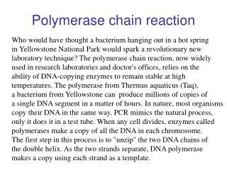
POLYMERASE CHAIN REACTION. PCR. Amplification of a specific DNA sequence (100-5000 bp) 2 synthetic oligonucleotide primers flanking the target sequence Use of thermostable DNA polymerase 3 step cycling process : -denaturation -annealing of primers -extension. Repeated cycle. history.

E N D
PCR • Amplification of a specific DNA sequence (100-5000 bp) • 2 synthetic oligonucleotide primers flanking the target sequence • Use of thermostable DNA polymerase • 3 step cycling process : -denaturation -annealing of primers -extension Repeated cycle
history • Kary Mullis • 1st thermal cycler :1986 • 3 waterbaths with different temperature, non thermostable DNA polymerase • Polymerase from Thermus aquaticus (Taq polymerase) : Topt:72C,withstand heating to 100C • Other polymerase: Pfu
DENATURATION • Denaturation: heating DNA to temperature above the Tm to make ssDNA. • if the DNA is heated in buffers of ionic strength lower than 150mM NaCl, the melting temperature is generally less than 100oC - which is why PCR works with denaturing temperatures of 91-97oC. • Taq polymerasehas ahalf-life of 30 min at 95oC, one should not do more than about 30 amplification cycles: however, it is possible to reduce the denaturation temperature after about 10 rounds of amplification, as the mean length of target DNA is decreased: for templates of 300bp or less, denaturation temperature may be reduced to as low as 88oC for 50% (G+C) templates (Yap and McGee, 1991), which means one may do as many as 40 cycles without much decrease in enzyme efficiency.
DENATURATION • "Time at temperature" is the main reason for denaturation / loss of activity of Taq: thus, if one reduces this, one will increase the number of cycles that are possible, whether the temperature is reduced or not. • Normally the denaturation time is 1 min at 94oC: it is possible, for short template sequences, to reduce this to 30 sec or less. • Increase in denaturation temperature and decrease in time may also work: Innis and Gelfand (1990) recommend 96oC for 15 sec.
annealing • T melting : depend on Primer length and sequence, the melting temperature of a DNA duplex increases with its length and (G+C) content • a simple formula for calculation of the primers Tm is Tm = 4(G + C) + 2(A + T)oC • annealing temperature (Ta) about 5oC below the lowest Tm of the pair of primers to be used (Innis and Gelfand, 1990) • if the Ta is increased by 1oC every other cycle, specificity of amplification and yield of products <1kb in length are both increased
annealing • If a Ta is too low : one or both primers willanneal to sequences other than the true target, as internal single-base mismatches or partial annealing may be tolerated • this is fine if one wishes to amplify similar or related targets; however, it can lead to "non-specific" amplification and consequent reduction in yield of the desired product
annealing • too high Ta : too little product will be made, as the likelihood of primer annealing is reduced • a pair of primers with very different Tas may never give appreciable yields of a unique product, and may also result in inadvertent "asymmetric" or single-strand amplification of the most efficiently primed product strand. • Annealing does not take long: most primers will anneal efficiently in 30 sec or less, unless the Ta is too close to the Tm, or unless they are unusually long
An illustration of the effect of annealing temperature on the specificity and on the yield of amplification of Human papillomavirus type 16 (HPV-16) (Williamson and Rybicki, 1991: J Med Virol 33: 165-171) Plasmid and biopsy sample DNA templates were amplified at different annealing temperatures as shown: note that while plasmid is amplified from 37 to 55oC, HPV DNA is only specifically amplified at 50oC.
EXTENSION • normally 70 - 72oC, for 0.5 - 3 min • elongation occurs from the moment of annealing,at around 70oC the activity is optimal, andprimer extension occurs at up to 100 bases/sec • About 1 min is sufficient for reliable amplification of 2kb sequences (Innis and Gelfand, 1990). Longer products require longer times: 3 min is a good bet for 3kb and longer products. Longer times may also be helpful in later cycles when product concentration exceeds enzyme concentration (>1nM), and when dNTP and / or primer depletion may become limiting.
REACTION BUFFER Recommended buffers generally contain : • 10-50mM Tris-HCl pH 8.3, • up to 50mM KCl, 1.5mM or higher MgCl2, • primers 0.2 - 1uM each primer, • 50 - 200uM each dNTP, • gelatin or BSA to 100ug/ml, • and/or non-ionic detergents such as Tween-20 or Nonidet P-40 or Triton X-100 (0.05 - 0.10% v/v) (Innis and Gelfand, 1990). Modern formulations may differ considerably
REACTION BUFFER • Higher than 50mM KCl or NaCl inhibits Taq, but some is necessary to facilitate primer annealing • [Mg2+] affects primer annealing; Tm of template, product and primer-template associations; product specificity; enzyme activity and fidelity. Taq requires free Mg2+, so allowances should be made for dNTPs, primers and template, all of which chelate and sequester the cation; of these, dNTPs are the most concentrated, so [Mg2+] should be 0.5 - 2.5mM greater than [dNTP]. A titration should be performed with varying [Mg2+] with all new template-primer combinations, as these can differ markedly in their requirements, even under the same conditions of concentrations and cycling times/temperatures.
REACTION BUFFER • Some enzymes do not need added protein, others are dependent on it. Some enzymes work markedly better in the presence of detergent, probably because it prevents the natural tendency of the enzyme to aggregate. • Primer concentrations should not go above 1uM unless there is a high degree of degeneracy; 0.2uM is sufficient for homologous primers. • Nucleotide concentration need not be above 50uM each: long products may require more, however.
Cycle Number • The number of amplification cycles necessary to produce a band visible on a gel depends largely on the starting concentration of the target DNA • Innis and Gelfand (1990) recommend from 40 - 45 cycles to amplify 50 target molecules, and 25 - 30 to amplify 3x10⁵ molecules to the same concentration. • This non-proportionality is due to a so-called plateau effect, which is the attenuation in the exponential rate of product accumulation in late stages of a PCR, when product reaches 0.3 - 1.0 nM. This may be caused by degradation of reactants (dNTPs, enzyme); reactant depletion (primers, dNTPs - former a problem with short products, latter for long products); end-product inhibition (pyrophosphate formation); competition for reactants by non-specific products; competition for primer binding by re-annealing of concentrated (10nM) product (Innis and Gelfand, 1990).
If desired product is not made in 30 cycles, take a small sample (1ul) of the amplified mix and re-amplify 20-30x in a new reaction mix rather than extending the run to more cycles: in some cases where template concentration is limiting, this can give good product where extension of cycling to 40x or more does not
nested primer PCR • PCR amplification is performed with one set of primers, then some product is taken - with or without removal of reagents - for re-amplification with an internally-situated, "nested" set of primers. This process adds another level of specificity, meaning that all products non-specifically amplified in the first round will not be amplified in the second
This gel photo shows the effect of nested PCR amplification on the detectability of Chicken anaemia virus (CAV) DNA in a dilution series: the PCR1 just detects 1000 template molecules; PCR2 amplifies 1 template molecule (Soiné C, Watson SK, Rybicki EP, Lucio B, Nordgren RM, Parrish CR, Schat KA (1993) Avian Dis 37: 467-476).
Helix Destabilisers / Additives With NAs of high (G+C) content, it may benecessary to use harsher denaturation conditions. For example, one may incorporate up to 10% (w or v/v) : • dimethyl sulphoxide (DMSO), • dimethyl formamide (DMF), • urea • or formamide These additives are presumed to lower the Tm of the target NA, although DMSO at 10% and higher is known to decrease the activity of Taq by up to 50% (Innis and Gelfand, 1990; Gelfand and White, 1990). Additives may also be necessary in the amplification of long target sequences:DMSO often helps in amplifying products of >1kb. Formamide can apparently dramatically improve the specificity of PCR (Sarkar et al., 1990), while glycerol improves the amplification of high (G+C) templates (Smith et al., 1990) Polyethylene glycol (PEG) may be a useful additive when DNA template concentration is very low: it promotes macromolecular association by solvent exclusion, meaning the pol can find the DNA
A simple set of rules for primer sequence design (adapted from Innis and Gelfand, 1991): • primers should be 17-28 bases in length; • base composition should be 50-60% (G+C); • primers should end (3') in a G or C, or CG or GC: this prevents "breathing" of ends and increases efficiency of priming; • Tms between 55-80oC are preferred; • runs of three or more Cs or Gs at the 3'-ends of primers may promote mispriming at G or C-rich sequences (because of stability of annealing), and should be avoided; • 3'-ends of primers should not be complementary (ie. base pair), as otherwise primer dimers will be synthesised preferentially to any other product; • primer self-complementarity (ability to form SECONDARY structures such as hairpins) should be avoided.
PRIMER DESIGN • Requires knowledge of some sequenceinformation • Gives high specificity and sensitivity • Allows amplification from limited startingmaterial
APPLICATION OF PCR • DIAGNOSTIC • BIODIVERSITY ANALYSIS • LABORATORY ROUTINE
Diagnostic Applications of PCR • detecting pathogens using genome-specific primer pairs • screening specific genes for unknown mutations • genotyping using known STS (Sequence Tagged Sites)markers
Laboratory applications of PCR • subcloning DNA targets using PCR, -T/A Cloning -Restriction Site Addition -Blunt-end Ligation • PCR-mediated in vitromutagenesis.
Reverse transcription polymerase chain reaction • Amplification from mRNA template • RNA strand is first reverse transcribed into its DNA complement (complementary DNA, or cDNA) using the enzyme reverse transcriptase, and the resulting cDNA is amplified using traditional PCR
The two-step RT-PCR process for converting RNA to DNA and the subsequent PCR amplification of the reversely-transcribed DNA: First strand reaction: complementary DNA (cDNA) is made from an mRNA template using dNTPs & reverse transcriptase. The components are combined with a DNA primer in a reverse transcriptase buffer for an hour at 42°C. Second strand reaction: after the reverse transcriptase reaction is complete, cDNA has been generated from the original ss mRNA, standard PCR (called the “second strand reaction”) is initiated.
In the two-step RT-PCR a thermostable DNA polymerase & the upstream and downstream DNA primers are added. Heating the reaction to temperatures above 37°C facilitates binding of DNA primers to the cDNA, & subsequent higher temperatures allow the DNA polymerase to make double-stranded DNA from the cDNA. Heating the reaction to ~95°C melts the two DNA strands apart, enabling the primers to bind again at lower temperatures and begin the chain reaction again. After ~30 cycles, millions of copies of the sequence of interest are generated
The use of RT-PCR • the diagnosis of genetic diseases • the determination of the abundance of specific different RNA molecules within a cell or tissue as a measure of gene expression • the cloning of eukaryotic genes in prokaryotes most eukaryotic genes contain introns which are present in the genome but not in the mature mRNA, the cDNA generated from a RT-PCR reaction is the exact DNA sequence which would be directly translated into protein after transcription. When these genes are expressed in prokaryotic cells such as E. coli for protein production/purification, the RNA produced directly from transcription need not undergo splicing as the transcript contains only exons (prokaryotes lack the mRNA splicing mechanism of eukaryotes) • studying the genomes of viruses whose genomes are composed of RNA, such as retroviruses like HIV
Real-time polymerase chain reaction • amplify and simultaneously quantify a targeted DNA molecule. measurement of DNA amplification during PCR in real time, i.e., the amplified product is measured at each PCR cycle • Two common methods of quantification are: (1) the use of fluorescent dyes that intercalate with double-stranded DNA, and (2) modified DNAoligonucleotide probes that fluoresce when hybridized with a complementary DNA • Frequently, real-time polymerase chain reaction is combined with reverse transcription to quantify messenger RNA (mRNA) in cells or tissues
Real-time PCR using double-stranded DNA dyes • A DNA-binding dye binds to all dsDNA in PCR, causing fluorescence of the dye. An increase in DNA product during PCR therefore leads to an increase in fluorescence intensity and is measured at each cycle, thus allowing DNA concentrations to be quantified. However, dsDNA dyes such as SYBR Green will bind to all dsDNA PCR products, including nonspecific PCR products (such as "primer dimers"). This can potentially interfere with or prevent accurate quantification of the intended target sequence • The reaction is run in a thermocycler, and after each cycle, the levels of fluorescence are measured with a detector; the dye only fluoresces when bound to the dsDNA (i.e., the PCR product). With reference to a standard dilution, the dsDNA concentration in the PCR can be determined
SYBR Green can’t bind single stranded DNA or primer But it can bind double stranded DNA once the polymerase has made the 2nd strand
Fluorescent reporter probe method • uses a sequence-specific RNA or DNA-based probe to quantify only the DNA containing the probe sequence; therefore, use of the reporter probe significantly increases specificity • allows quantification even in the presence of some non-specific DNA amplification • allows for multiplexing - assaying for several genes in the same reaction by using specific probes with different-coloured labels, provided that all genes are amplified with similar efficiency
commonly carried out with an RNA-based probe with a fluorescent reporter at one end and a quencher of fluorescence at the opposite end of the probe. The close proximity of the reporter to the quencher prevents detection of its fluorescence; breakdown of the probe by the 5' to 3' exonuclease activity of the taq polymerase breaks the reporter-quencher proximity and thus allows unquenched emission of fluorescence, which can be detected. An increase in the product targeted by the reporter probe at each PCR cycle therefore causes a proportional increase in fluorescence due to the breakdown of the probe and release of the reporter
The PCR is prepared as usual and the reporter probe is added. As the reaction commences, during the annealing stage of the PCR both probe and primers anneal to the DNA target. Polymerisation of a new DNA strand is initiated from the primers, and once the polymerase reaches the probe, its 5'-3-exonuclease degrades the probe, physically separating the fluorescent reporter from the quencher, resulting in an increase in fluorescence. Fluorescence is detected and measured in the real-time PCR thermocycler, and its geometric increase corresponding to exponential increase of the product is used to determine the threshold cycle (CT) in each reaction.
In intact probes, reporter fluorescence is quenched. • (2) Probes and the complementary DNA strand are hybridized and reporter fluorescence is still quenched. • (3) During PCR, the probe is degraded by the Taq polymerase and the fluorescent reporter released.
Principle of quantitation • The number of cycles it takes to reach a certain amount of fluorescence is proportional to the amount of cDNA present at the start • This can be plotted as cycle no v log concentration to give a straight line • By using known amounts of cDNA you can obtain a standard curve to find out levels of an unknown
The use of real time PCR • rapidly detect the presence of genes involved in infectious diseases, cancer and genetic abnormalities • determining how the genetic expression of a particular gene changes over time, such as in the response of tissue and cell cultures to an administration of a pharmacological agent, progression of cell differentiation, or in response to changes in environmental conditions • used in environmental microbiology, for example to quantify resistance genes in water samples