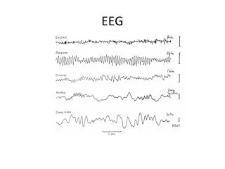
Classic EEG Abnormalities
300 likes | 477 Views
Classic EEG Abnormalities. Academic Half-Day June 5th 2013. How do you read an eeg ?. How can an eeg be abnormal?. Epileptic Focal or Generalized Interictal Focal or Generalized Seizure Non-convulsive Status Periodic PLEDs/ BiPLEDs GPEDs Burst-suppression Triphasic waves

Classic EEG Abnormalities
E N D
Presentation Transcript
Classic EEG Abnormalities Academic Half-Day June 5th 2013
Epileptic • Focal or Generalized Interictal • Focal or Generalized Seizure • Non-convulsive Status • Periodic • PLEDs/BiPLEDs • GPEDs • Burst-suppression • Triphasic waves • Periodic complexes (CJD) • Background Abnormality
Case One • How do you decide it is epileptic activity? (inter-ictal) • Sharp, asymmetrical (rapid rise) • Voltage maximum • “Field” • Slow wave • Recurs Fisch and Spehlman’s EEG Primer
Case Two • It is generalized • It is “inter-ictal” • There is normal background
Case Three • “it is what it sounds like” • No normal background • It is periodic (which means…) • It is bilateral
Case Four • It looks epileptic • Generalized • “Neat and orderly” • Normal background • Don’t get thrown off by high amplitude
Case Five • “it is what it sounds like” • Epileptic, Periodic, Lateralized • Don’t worry about volume conduction Chong DJ and Hirsch LJ. Which EEG patterns warrant treatment in the critically ill?Reviewing the evidence for treatment of periodic epileptiform discharges and related patterns. J ClinNeurophysiol 2005;22:79.
Case Six • Repetitive spikes or sharp waves (alone or in complexes with slow waves) at > 2.5 / sec • Above, < 2.5 / sec, with either clinical ictal phenomena or response to AED • Rhythmic slow waves with evolution in frequency or location Kaplan P. EEG criteria for non-convulsive status epilepticus. Epilepsia 2007; 48 (suppl 8):39-41.
Case Seven • Generalized • Synchronous • Periodic Foreman et al. Generalized periodic discharges in the critically ill. A case control study of 200 patients. Neurology 2012;79:1951-60.
Case Eight • Focal or generalized • Looks epileptic (sharp) • Continuous
Case Nine • Fairly strict criteria; it does have to be periodic and triphasic • But…mimics • So…clinical context Kaplan P. EEG criteria for non-convulsive status epilepticus. Epilepsia 2007; 48 (suppl 8):39-41.
Case Ten • Periodic • Clinical context • “The least unique” • Usually bilateral but can be unilateral • Sharp waves but variable morphology Weiser et al. EEG in Creutzfeld-Jakob disease. ClinNeurophysiol 2006; 117: 935-51.