Download
1 / 115
1.33k likes | 1.97k Views
HEART FAILURE IN NEONATE AND INFANT. DR RAJESH KF. Congestive heart failure (CHF) refers to a clinical state of systemic and pulmonary congestion resulting from inability of the heart to pump as much blood as required for the adequate metabolism of the body.

E N D
HEART FAILURE IN NEONATE AND INFANT DR RAJESH KF
Congestive heart failure (CHF) refers to a clinical state of systemic and pulmonary congestion resulting from inability of the heart to pump as much blood as required for the adequate metabolism of the body. • Clinical picture of CHF results from a combination of “relatively low output” and compensatory responses to increase it
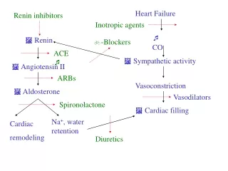
PATHOPHYSIOLOGY Unmet tissue demands for cardiac output result in activation of • Renin-aldosteroneangiotensin system • Sympathetic nervous system • Cytokine-induced inflammation • “signaling” cascades that trigger cachexia.
Longstanding increases in myocardial work and myocardial oxygen consumption (MVO2) ultimately worsen HF symptoms and lead to a chronic phase that involves cardiac remodeling
CARDIACREMODELING? • Maladaptive cardiac hypertrophy • Expansion of the myofibrillar components of individual myocytes (new cells rarely form) • An increase in the myocyte/capillary ratio • Activation and proliferation of abundant nonmyocytecardiac cells, some of which produce cardiac scarring • Produce a poorly contractile and less compliant heart
Endogenous mechanisms defend progressive HF • Stimulation of insulin like growth factor and GH • ANP and BNP are hormones secreted by the heart in response to volume and pressure overload that increase vasodilation and diuresis acutely and chronically prevent inflammation, cardiac fibrosis and hypertrophy.
CLINICAL MANIFESTATIONS IN INFANTS WITH HF • Variety of age dependent clinical presentations • In neonates, the earliest clinical manifestations may be subtle
CLINICAL MANIFESTATIONS IN INFANTS WITH HF • Feeding difficulties • Rapid respirations • Tachycardia • Cardiacenlargement • Gallop rhythm (S3) • Hepatomegaly • Pulmonary rales • Peripheral edema • Easy fatigability. • Sweating • Irritability • failure to thrive.
Feeding difficulties & increased fatigability Important clue in detecting CHF in infants • Often it is noticed by mother • Interrupted feeding (suck- rest -suck cycles) • Infant pauses frequently to rest during feedings • Inability to finish the feed, taking longer to finish each feed (> 30 minutes) • Forehead sweating during feeds –due to activation of sympathetic nervous system –a very useful sign • Increasing symptoms during and after feedings
Rapid respirations • Tachypnea > 60/min in 0-2mth >50/mt in 2mth to 1yr >40/mt 1-5 yr in calm child • Happy tachypnea- tachypnea with out much retractions • Grunting (a form of positive end-expiratory pressure) • In cyanotic heart disease rapid respirations may be due to associated brain anoxia and not CHF -treatment for these two conditions is entirely different • Fever especially with a pulmonary infection may produce rapid respirations.
Tachycardia • Rate is difficult to evaluate in a crying or moving child • Tachycardia in the absence of fever or crying when accompanied by rapid respirations and hepatomegaly is indicative of HF • Persistently raised heart rate > 160 bpm in infants • > 100 bpm in older children. • Consider SVT if heart rate > 220 bpm in infants and > 180 bpm in older children.
Cardiomegaly • Consistent sign of impaired cardiac function, secondary to ventricular dilatation and/or hypertrophy. • May be absent in early stages, especially with myocarditis, arrhythmias, restrictive disorders and pulmonary venous obstruction(obstructed TAPVC) • Apex 4th space 1cm outside MCL in newborn
Hepatomegaly • Lower edge of the liver is palpable 1 to 2 cms below right costal margin normally in infancy • In the presence of respiratory infection increased expansion of the lungs displace liver caudally • Usually in such circumstances the spleen is palpable • Hepatomegaly is a sign of CHF • Decrease in size is an excellent criterion of response to therapy
Pulmonary rales • Of not much use in detecting CHF in infants • Rales may be heard at both lung bases • When present are difficult to differentiate from those due to the pulmonary infection which frequently accompanies failure
Peripheral edema • Edema is a very late sign of failure in infants and children • Presacral and posterior chest wall edema in young infants • It indicates a very severe degree of failure. • Daily wtmonitoring is useful in neonates -- rapid increase in wt > 30 gm /day may be a clue to CCF and is useful in monitoring response to treatment.
Cold extremity, low blood pressure, skin mottling are signs of impending shock • Pulsusalternans (alternate strong and weak contractions of a failing myocardium),or pulsusparadoxus (decrease in pulse volume and blood pressure with inspiration) are frequently observed in infants with severe CHF
CLASSIFICATION • NYHA Heart Failure Classification is not applicable • Ross Heart Failure Classification was developed for global assessment of heart failure severity in infants • Modified to apply to all pediatric ages • Modified Ross Classification incorporates Feeding difficulties Growth problems Symptoms of exercise intolerance
MODIFIED ROSS HEART FAILURE CLASSIFICATION FOR CHILDREN Class I • Asymptomatic Class II • Mild tachypnea or diaphoresis with feeding in infants • Dyspnea on exertion in older children Class III • Marked tachypnea or diaphoresis with feeding in infants • Marked dyspnea on exertion • Prolonged feeding times with growth failure Class IV • Symptoms such as tachypnea, retractions, grunting, or diaphoresis at rest
The time of onset of CHF holds the key to the etiological diagnosis in this age group
Parallel circulation becomes series at birth • Cardiac anomalies present at that point are • Critical AS • HLHS • Mitral atresia
Functional closure PDA 1 to 2weeks • PDA dependent lesions ,depend on patent duct for either • pulmonary blood flow-Fallots with pulmonary atresia • systemic blood flow-IAA/COA • mixing of systemic and pulmonary blood-TGA • Present at 1 to 2weeks
Anatomic closure of PDA by 2to4 weeks • Coarctation of aorta
Pulmonary vascular resistance falls 4to 6weeks • Congestive heart failure due to L-R shunt • Large VSD • PDA • ALCAPA
CHF in the fetus • Disorders that are fatal in the immediate neonatal period are often well tolerated in the fetus due to the pattern of fetal blood flow (e.g. TGA) Causes of CHF in the fetus • SVT • Severe bradycardiadue to CHB • Anemia • Severe TR due to Ebstein’s anomaly or MR from AV canal defect • Myocarditis
Most of these are recognized by fetal echo • Severe CHF in the fetus produces hydropsfetalis with ascites, pleural and pericardial effusions and anasarca. • Digoxin or sympathomimetics to the mother may be helpful in cases of fetal tachyarrhythmia or CHB respectively.
Premature neonates PDA • poor myocardial reserve • Fluid overload
CHF on first day of life • Myocardial dysfunction secondary to asphyxia, hypoglycemia, hypocalcaemia or sepsis are usually responsible for CHF on first day • Few structural heart defects cause CHF within hours of birth • HLHS, severe TR or PR, Large AV fistula • TR secondary to hypoxia induced papillary muscle dysfunction or Ebstein’s anomaly of the valve • Improves as the pulmonary artery pressure falls over the next few days
CHF in first week of life • Serious cardiac disorders which are potentially curable but carry a high mortality if untreated often present with CHF in the first week of life • A sense of urgency should always accompany evaluation of the patient with CHF in the first week • Closure of the ductusarteriosus is often the precipitating event • Prostaglandins E1 should be utilised
Peripheral pulses and oxygen saturation (pulse oximeter) should be checked in both the upper and lower extremities A lower saturation in the lower limbs means right to left ductal shunting due to PAH or AAI • ASD or VSD does not lead to CHF in the first two weeks of life, an additional cause must be sought (eg.COA or TAPVC).
TGA no VSD -1ST week VSD and no PS-6-8 weeks • Critical AS or PS • Obstructive TAPVC • Adrenal insufficiency due to enzyme deficiencies or neonatal thyrotoxicosis could present with CHF in the first few days of life
ALPROSTODIL • Prostaglandins E1 • Maintain patency of ductus • Cyanotic lesions TGA • LT sided obstructive lesions HLHS, critical AS,COA,IAA • Available as inj500microgm/ml • IV 0.05 to0.1microgm /kg/min • 0.01 to 0.05 microgm /kg/min maintainance • Vasodilation of all arteries including ductus
Monitor spo2,RR, HR,BP,ECG,temp Complications • apnea, • Seizure • Hypotension • Bradycardia • Tachycardia • cardiac arrest • fever • Extravasation may cause sloughing and necrosis
CHF beyond second week of life • Most common cause of CHF in infants is VSD • Presents around 6-8 weeks of age. • Left to right shunt increases as the PVR falls • Murmur of VSD is apparent by one week • Full blown picture of CHF occurs around 6-8 weeks. • Other left to right shunts like PDA present similarly • Fall in PVR is delayed in presence of hypoxic lung disease and at high altitude and can alter the time course • Spontaneous improvement in CHF -development of obstructive pulmonary arterial hypertension even in early childhood
ALCAPA a rare disease in this age group • It is curable • As the pulmonary artery pressure decreases in the neonatal period, these babies suffer from episodes of excessive crying with sweating (angina) and myocardial infarction. • ECG shows pathologic q waves • Often misdiagnosed as having “dilated cardiomyopathy”
CAUSES OF HF IN CHILDREN CARDIAC Congenital structural malformations ● Excessive Preload ● Excessive Afterload ● Complex congenital heart disease No structural anomalies ● Cardiomyopathy ● Myocarditis ● Myocardial infarction ● Acquired valve disorders ● Hypertension ● Kawasaki syndrome ● Arrhythmia (bradycardia or tachycardia) NONCARDIAC ● Anemia ● Sepsis ● Hypoglycemia ● Diabetic ketoacidosis ● Hypothyroidism ● Other endocrinopathies ● Arteriovenous fistula ● Renal failure ● Muscular dystrophies
VOLUME OVERLOAD (EXCESSIVE PRELOAD) • Left-to-right shunting VSD PDA AP window AVSD ASD(rare) Total/Partial Anomalous Pulmonary Venous Connection • AV or semilunar valve insufficiency AR in bicommissural aortic valve/after valvotomy MR after repair of AVSD PR after repair of TOF Severe TR in Ebstein anomaly
Right-sided volume loading • Large ASD or anomalous pulmonary vein connections • Congenital or surgically acquired PR especially if downstream pulmonary arterial narrowing • Highly compliant RV accepts significant volume -without increasing filling pressure • Rarely causes HF early in life
PRESSURE OVERLOAD (EXCESSIVE AFTERLOAD) • Left sided obstruction Congenital AS Aortic coarctation Lethal arrhythmias - severe afterload stress? ?HTN • Right-sided obstruction Severe PS
Left heart obstructive lesions • First postnatal week-ductusarteriosuscloses • Increased LVEDP and a decreased pressure gradient between the aorta and ventricle at end-diastole produce subendocardialischemia due to inadequate coronary flow • Increased afterload and subendocardial ischemia result in HF syndrome
COMPLEX CONGENITAL HEART DISEASE Abnormal RV • CCTGA • D TGA Single ventricle physiology • HLHS • Unbalanced AVSD • Post Fontan procedure
Often combined volume and pressure overload • Both systemic and pulmonary circulations can be affected • Cyanosis in CCHD-risk of subendocardial ischemia contributing to impaired ventricular performance • Molecular abnormalities in transcription factors that lead to congenital structural abnormalities – also associated with abnormal myocardial performance and arrhythmias
ABNORMAL RV • In pediatric heart disease much of the pathology is due to an abnormal RV • RV myocytes appear to be structurally identical to LV myocytes • Differences in contraction compared to the LV are due to the shape of the RV and myocardial organization
Gene expression patterns are different in the RV and the LV, which may affect function. • Genes that affect angiotensin and adrenergic receptor signaling showed lower expression in the RV than the LV • Genes that contribute to maladaptive signaling showed higher expression in the RV
Hypoplastic right heart syndromes -3 parts of the RV do not form normally or may be missing entirely. • Defects in the IVS or abnormal LV function-Adversely affect the third phase of normal RV contraction through its interdependence on normal septal function
Volume overload of the RV • Can arise through significant PR or TR • Compensatory dilation to decompensated dilation occur slowly
Increased RV afterload • RVOT obstruction • RV serving as the systemic ventricle • Usually can adapt if present at birth • Once the RV assumes a mature, thin-walled configuration, it cannot always mount a hypertrophic response • RV is able to support the systemic circulation for many years but function often deteriorates over time
SINGLE VENTRICLE PHYSIOLOGY • Ventricular morphology (left, right, indeterminate, or unbalanced) results in a single functional pumping chamber • At birth presentation depends on the morphology • Range from well-tolerated cyanosis to decompensated heart failure and cardiogenic shock • double inlet ventricle(SV), HLHS , Tricuspid atresia, isomerism