Download
1 / 21
520 likes | 1.35k Views
Lung Cancer. By Dhara Mehta, 1068. Introduction to Lung Cancer. Lung cancer is the uncontrolled growth of abnormal cells in one or both lungs. As they grow, the abnormal cells can form tumors and interfere with the functioning of the lung, which provides oxygen to the body via the blood .

E N D
Lung Cancer By Dhara Mehta, 1068
Introduction to Lung Cancer • Lung cancer is the uncontrolled growth of abnormal cells in one or both lungs. • As they grow, the abnormal cells can form tumors and interfere with the functioning of the lung, which provides oxygen to the body via the blood. • They arise from epithelial cells
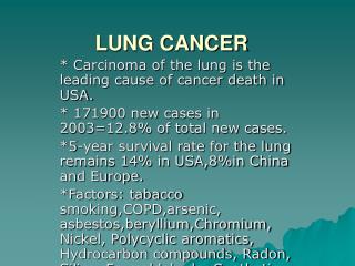
Epidemiology • Worldwide, lung cancer is the most common cancer in terms of both incidence and mortality. • The highest rates are in Europe and North America • Higher chances of lung cancer in people aged over 50 and who have a history of smoking. • There was higher mortality in men, but now women’s mortality rate is increasing.
Risk factors/ Causes • the most common cause is long-term exposure to tobacco smoke • attributed to a combination of genetic factors (gene mutations), and exposure to; radon gas,asbestos,and air pollutionincluding second-hand smoking. • Family history
Classification of Lung Cancer • There are two major subdivisions of lung cancer • NSCLC- Non Small Cell Lung Carcinoma (85%) • Squamous Cell Carcinoma- 25-30% • Adenocarcinoma- 40% • Large Cell Carcinoma- 10-15% • SCLC- Small Cell Lung Carcinoma- 15%
Squamous Cell Carcinoma • More in Males and smokers • central (hilar) location • They typically occur close to large airways. • forms in the lining of the bronchial tubes. • accounts for about 30% • P53 mutation is common in SCC
Adenocarcinoma • Nearly 40% of lung cancers are adenocarcinoma, which usually originates in peripheral lung tissue • Affects women, non smokers and smokers also (most common) • Occurs at a younger age (<40yrs) • See Kras mutation (mainly in smokers) • EGFR-3 mutations in non smokers and women • Slow growing and discovered on the outer area of lung • Types: acinar, solid, papillary, broncho-alveolar, mucin forming,mixed Mucinous Adenocarcinoma
Large Cell Carcinoma • Called so because the cancer cells are large, with excess cytoplasm and large nuclei • Poor prognosis and early metastasis Large cell Carcinoma
SCLC- Small Cell Lung Carcinoma • See mainly in males • Related to smoking • Grows more rapidly • More responsive to chemo • Cells resemble lymphocytes • Inconspicuous nucleoli • Vascular invasion • Infiltrate and metastasize widely • Neuro endocrine orgins (paraneolastic syndromes)-cells contain dense neurosecretory granules • See p53 and RB1 mutations SCLC
Signs and Symptoms • may not appear until the disease is advanced. • respiratory symptoms: coughing, coughing up blood, wheezing, shortness of breath, change in color or sputum, recurrent lung problems • systemic symptoms: weight loss, fever, clubbing of the fingernails, fatigue, neurological symptoms • symptoms due to local compression: chest pain, bone pain, superior vena cava obstruction, difficulty swallowing • Common sites of spread include the brain, bone, adrenal glands, opposite lung, liver, pericardium, and kidneys.
Diagnosis • Performing an x-ray is one of the first steps that may suggest lung cancer. This may reveal mass, atelectasis (collapse), consolidation (pneumonia), or pleural effusion. • Sputum cytology- reveal the presence of lung cancer cells. • CT imaging is typically used to provide more information about the type and extent of disease. • Bronchoscopy or CT-guided biopsy is often used to sample the tumor for histopathology.
Staging of Lung Cancer • Stage I: The cancer is located only in the lungs and has not spread to any lymph nodes. • Stage II: The cancer is in the lung and nearby lymph nodes. • Stage III: Cancer is found in the lung and in the lymph nodes in the middle of the chest, also described as locally advanced disease. Stage III has two subtypes: • If the cancer has spread only to lymph nodes on the same side of the chest where the cancer started, it is called stage IIIA. • If the cancer has spread to the lymph nodes on the opposite side of the chest, or above the collar bone, it is called stage IIIB. • Stage IV: This is the most advanced stage of lung cancer, and is also described as advanced disease. Cancer has spread to both lungs, to fluid in the area around the lungs, or to another part of the body.
Management of NSCLC • Treatment depends on the cancer's specific cell type, how far it has spread, and the person's performance status. • Surgery • Most stage I and stage II. Surgeon removes the lobe, or section, of the lung containing the tumor. • Some surgeons use video-assisted thoracoscopic surgery (VATS) • https://www.youtube.com/watch?v=KoBw_-xj68E
Chemotherapy and Radiation • Evidence suggests that chemotherapy after surgery may help prevent the cancer from returning. • For people with stage III and IV lung cancer that cannot be removed surgically, chemotherapy in combination with definitive (high-dose) radiation treatments. In stage IV patients, radiation is used only for palliation of symptoms. • consists of a combination of drugs such as cisplatin(Platinol) or carboplatin (Paraplatin) plus docetaxel (Taxotere), gemcitabine (Gemzar), paclitaxel (Taxol and others), vinorelbine (Navelbine and others), or pemetrexed (Alimta). • the lung cancer may come back. Doctors will prescribe a second course of drug treatment referred to as second-line chemotherapy.
Targeted Treatments • Targeted therapies are designed specifically to attack cancer cells by attaching to or blocking targets that appear on the surfaces of those cells. • People who have advanced lung cancer with certain molecular biomarkers may receive treatment with a targeted drug alone or in combination with chemotherapy. These treatments for lung cancer include: • Erlotinib (Tarceva)–blocks the epidermal growth factor receptor (EGFR). EGFR allowing substances in that encourage a cancer cell to grow and spread. • Bevacizumab(Avastin)- blocks vascular endothelial growth factor-VEGF so new blood vessels can’t grow
Management: SCLC • Chemotherapy and Radiation Therapy • chemotherapy is an essential part of treatment. Radiation treatment may be used as well depending on the stage of cancer. • Preventive Radiation Therapy to the Brain • helps prevent the cancer from spreading to the brain. This procedure is known as prophylactic cranial irradiation (PCI). • Surgery- rarelyusedbecauseit is fastgrowing
Complications • Shortness of breath • Coughing up blood. • Fluid in the chest (pleural effusion)- Lung cancer can cause fluid to accumulate in the space that surrounds the affected lung in the chest cavity (pleural space). • Cancer that spreads to other parts of the body (metastasis)can cause pain, nausea, headaches, or other signs and symptoms depending on what organ is affected.
Prognosis • 1-yr relative survival rate 41% • According to the American Cancer Society, patients diagnosed with stage I or stage II NSCLC have a 30-49 percent five-year survival rate • SCLC is far more aggressive. If not treated, the median survival rate is two to four months, according to The National Cancer Institute.
Case Study • A 20-year-old man with no history of tobacco use presented with a several-months’ history of cough and lower back pain, and an 11.3-kg weight loss. Because of the persistent cough and development of hemoptysis, further imaging studies were obtained. A chest radiograph revealed total opacification of the right lung Posteroanterior view of the chest demonstrates complete opacification of the right hemithorax.
Case Study, cont. • Computed tomography imaging of the thorax revealed a 7×7×8- cm mass in the superior right hilum. • Further imaging revealed retroperitoneal lymphadenopathy, renal and pancreatic masses, skeletal metastases. • These findings confirmed a primary lung adenocarcinoma. https://www.youtube.com/watch?v=hy3lxAtyD_M
References • http://www.ncbi.nlm.nih.gov/pmc/articles/PMC2826778/ • http://www.lungcancer.org • http://www.medicinenet.com/lung_cancer/article.htm • http://www.nhs.uk/conditions/cancer-of-the-lung/Pages/Introduction.aspx • http://www.cancer.gov/cancertopics/types/lung • http://www.mayoclinic.org/diseases-conditions/lung-cancer/basics/treatment/con-20025531 • http://www.ncbi.nlm.nih.gov/pubmed/21642865 • http://www.healthline.com/health-slideshow/non-small-cell-lung-cancer-vs-small-cell#promoSlide