Download
1 / 10
100 likes | 208 Views
Learn about the pulmonary and systemic circuits of the circulatory system, the heart's control mechanisms, and blood flow patterns. Explore the closed and open circulatory systems and the role of the lymphatic system in maintaining fluid balance.

E N D
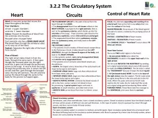
3.2.2 The Circulatory System Circuits THE PULMONARY CIRCUIT is the path of blood from the heart through the LUNGS. • The deoxygenated blood from all tissues collects in the RIGHT ATRIUM, is pumped to the right ventricle, then is sent to the pulmonary arteries, which divide up into the arterioles of the lungs. These arterioles take blood to the pulmonary capillaries, where CO2 and O2 are exchanged. • The oxygenated blood then enters pulmonary venules, then the pulmonary veins, and finally back to the LEFT ATRIUM. THE SYSTEMIC CIRCUIT • The systemic circuit includes all blood vessels except those in the Pulmonary Circuit. It takes blood from the LEFT VENTRICLE, through the tissues & organs of the body, and back to the RIGHT ATRIUM. • in the systemic system, veins carry deoxygenated blood, and arteries carry oxygenated blood. • The systemic circuit contains some blood vessels you should know: • AORTA: the largest artery. Branches of the aorta lead to all major body regions and organs. • SUPERIOR VENAE CAVA: large vein that collects blood from head, chest, and arms. • INFERIOR VENAE CAVA: large vein that collects blood from the lower body regions and organs. • HEPATIC PORTAL SYSTEM: connects the blood vessels of villi to the liver, carries nutrient rich blood to liver for processing. A portal system begins and ends in capillaries (in small intestine, and other end in liver. • HEPATIC VEIN carries blood from liver to inferior venae cava. Heart Control of Heart Rate PULSE: the alternate expanding and recoiling of an arterial wall that can be felt in any artery that runs near the surface of the body BLOOD PRESSURE: the pressure of the blood against the wall of a vessel, created by the pumping action of the heart. SYSTOLE = CONTRACTION of heart muscle. DIASTOLE = RELAXATION of heart muscle. The CARDIAC CYCLE (= “heartbeat”) occurs about 70 times per minute Heart Rate Control There are TWO nodal regions in the heart: 1. SA (sinoatrial) NODE (also called the PACEMAKER): located in the upper back wall of the right atrium. The SA node INITIATES THE HEARTBEAT by sending out a signal automatically about every 0.85 seconds to make the ATRIA CONTRACT. The SA node is called the “PACEMAKER” because it keeps the beat regular 2. AV (atrioventricular) NODE: found in the base of the right atrium near the septum. The SA node sends its signal along fibres to the atria and also to the AV node. When the pulse sent out by the SA node reaches the AV node, the AV node itself the sends out a signal along special conducting fibres called PURKINJE FIBRES. These fibres take the message to the VENTRICLES, and cause them to contract. Heart: A muscular pump that moves the blood throughout the body. Four chambers: atrium- 2 upper chambers ventricle- 2 lower chamber Valves: Prevent the backflow of blood from the ventricles into the atria Bicuspid valve: tricuspid Valve Each ventricle also has a SEMILUNAR VALVE The blood flows through the semilunar vales on its way out of the heart. Septum: Separates the right side of the heart from the left Flow of blood: The right atrium takes blood in from the body, through the vena cava’s. It then goes though the tricuspid valve into the right ventricle. It goes out through the pulmonary artery to the lungs. It returns from the lungs through the pulmonary vein. It enters the left atrium and then goes through the bicuspid valve into the left ventricle. The left ventricle pumps the blood through the aorta to the body for the process to happen again. Closed circulatory systemVertebrates, and a few invertebrates, have a closed circulatory system. Closed circulatory systems have the blood closed at all times within vessels of different size and wall thickness. In this type of system, blood is pumped by a heart through vessels, and does not normally fill body cavities. Open circulatory systemThe open circulatory system is common to molluscs and arthropods. Open circulatory pump blood into a hemocoel with the blood diffusing back to the circulatory system between cells. Blood is pumped by a heart into the body cavities, where tissues are surrounded by the blood.
3.2.3 The Blood Blood and Blood vessels The Lymphatic System • The lymphatic system is another vascular system in your body. It is separate from your cardiovascular system (i.e. it has its own veins and capillaries) but it is ultimately connected back with the cardiovascular system (i.e. the fluid from the lymphatic system eventually gets sent back into the bloodstream). The function of the lymphatic system is to take up excess tissue fluid from the tissues and returns it to the cardiovascular system. • It is a one-way system that starts in the tissues and empties into the cardiovascular system. • Lymph vessels consist of LYMPH CAPILLARIES and LYMPH VEINS (which have valves).The fluid in the lymph vessels it is called LYMPH. • Lymph contains LYMPHOCYTES which are a type of white blood cell. Some lymphocytes produce antibodies. Other Parts of the Lymphatic system you should know: • Lacteal: blind ends of lymph vessels in villi of the small intestine. Products of fat digestion enter here. The lacteals transport lipids to the blood. • LYMPH NODES: small oval or round structures that occur along strategic places on lymph vessels. They produce and store lymphocytes, and filter lymph of damaged cells and debris. • SPLEEN: located behind the stomach. Contains white blood cells and stores blood. • THYMUS GLAND: located in the upper thoracic cavity, functions in production and maturation of some lymphocytes. Decreases in size with age (may be a factor in aging). Summary of Main Functions of Lymphatic System 1. transport of excess tissue fluid back to cardiovascular system 2. absorption of fat from the intestine and transport to blood 3. fighting infection by production of lymphocytes Some lymphocytes produce antibodies. Blood-:plasma-liquid of blood 92% water and 8% protein Functions of the blood • Transport of O2, nutrients both organic such as :glucose, vitamins, amino acids, fatty acids and glycerol; and inorganic ions such as: Na+, K+, Cl-, Ca2+ and I- to all body cells • Transport of CO2 and wastes . It transports metabolic wastes such as : urea, creatinineand uric acid away from cells • Transport of chemical messengers inlcuding hormones to cells • Maintaining pH of body fluids • Distributing heat and maintaining body temperature • Maintaining water content and ion concentration of body fluids • Protection against disease-causing micro-organisms Platelets- These have no nucleus, they are fragments of cells that help the blood to clot. They are formed in red bone marrow. They release fibres to work with protein to create blood clots, which form plugs to help reduce blood loss. RBC- red blood cells- most abundant type of cells. They contain Haemoglobin, and are biconcave discs. They do not contain a nucleus. They transport oxygen which they get when it diffuses into the capillaries from the alveoli. They are made in red bone marrow. They are destroyed in liver and spleen. WBC- white blood cells. Their function is to defend against disease. They do this either by phagocytosis where the WBC engulf pathogens or by the production of antibodies. They are produced in yellow bone marrow. Plasma – the fluid part of the blood that contains dissolved substances Types of Blood Vessels Arteries: large blood vessels that carry oxygenated blood. They have small lumens and thick walls to withstand high blood pressure. They carry blood from the heart to the body. They have elastic walls. Veins: Blood vessels that carry deoxygenated blood from the body to the heart. They have thin walls and large lumens, They contain valves. Capillaries: Tiny blood vessels that absorb food and oxygen to feed RBC and get rid of CO2 and waste. They connect arteries to Veins.
3.5.3 The Eye and the Ear The Eye The Ear • The conjunctiva is the membrane surrounding the eye. It protects the eye. • The sclera is a tough, white coat that holds the eye in shape. • The cornea is the front part of the sclera. It allows light into the eye and bonds it to help focus light on the retina. • The choroid nourishes the eye and prevents internal reflection of light. • Theretina is light sensitive. It contains light receptors, rods (for black and white vision and to see in dim light) and cones (for colour vision, and to see in bright light). • The fovea is the part of the retina where most images are focused. • The blind spot is where the optic nerve leaves the retina. It has no rods or cones. • The optic nerve carries impulses to the brain. • The lens focuses light on the retina. • The iris is the coloured part of the eye. It controls the amount of light entering the eye. • The pupil is the black circle at the front of the eye. It lets light into the eye. • TheCiliary muscles change the shape of the lens (called accommodation) to focus the image on the retina. • The aqueous and vitreous humours is fluid that helps to keep the eye in shape. • The ear is the organ of hearing. • The pinna or ear lobe collects vibrations. • The auditory canal carries vibrations from the pinna to the eardrum. • The eardrum carries the vibrations to the middle ear. • The ossicles(the hammer, anvil and stirrup) increasethe amplitude of the vibrations and pass them on to the oval window. • The Eustachian tube, connects the middle ear with the pharynx and equalises pressure between the middle and outer ear. • The cochlea is responsible for hearing. It converts vibrations into electrical impulses that are sent to the brain along the auditory nerve. • The organ of Cortiin the cochlea contains receptor cells that us to hear • The semi-circular canals are responsible for balance.
3.5.3 The Nervous System The Nervous System Neurons A neuron is a nerve cell. There are three types of neurons. Sensory (afferent) neurons take messages to the CNS. Motor (efferent) neurons take messages from the CNS. Interneurons connect motor and sensory neurons in the CNS. The main parts of a neuron: Nerve endings connect to receptors or sense organs. Dendrites (can be long or short, and branched) carry impulses to a cell body. Axons carry nerve impulses away from cell bodies. Schwann cells make myelin. Myelin insulates the electrical impulses The cell body forms neurotransmitters and the different parts of the neuron. Neurotransmitters are chemical s released across a synaptic cleft to carry a signal from one neuron to another. The chemical is then destroyed or removed The central Nervous System is divided into 1 - Central Nervous System - including the brain & spinal cord 2 - Peripheral Nervous System - including cranial nerves, spinal nerves, & all branches of cranial & spinal nerves Nervous coordination involves: • reception (detecting a stimulus) • transmission (movement of the impulse) • integration (impulses are sorted, processed and decisions made) • response (a muscle or gland carries out the effect) Transmission of an Impulse • A synapse is the region where two neurons come into close contact. • A synaptic cleft is a tiny gap between one neuron and either another neuron or an effector. • Synapses control the direction of impulses. • A reflex action is an automatic response to a stimulus. • Reflex actions are fast responses that are designed to protect the body • A reflex arc is the basic unit of function of the nervous system. It consist of receptors, nerves and effectors. • The route taken along a reflex arc is: • receptor → sensory neuron → spinal cord → interneuron → motor neuron → effector. • At the interneuron stage an impulse is also sent to the brain. The brain is made aware of the action, but does not control it.
3.5.3 The Brain and Spinal Cord The Brain The Spinal Cord The spinal cord carries impulses to and from the brain and controls many reflex actions. In the spinal cord: sensory neurons enter through dorsal roots the dorsal root ganglion contains the cell bodies of the sensory neurons white matter contains axons (mostly) grey matter contains cell bodies and dendrites (mostly) interneurons connect sensory and motor neurons motor neurons emerge through ventral roots The brain is protected by the skull bones, meninges (threemembranes) and cerebrospinal fluid. The cerebrum is our conscious brain, with different parts havingdifferent jobs to do e.g. memory, intelligence, personality. The hypothalmus(osmoregulation/homoestasis) is the centre for the regulation of the internalorgans The pituitary ‘[master] gland secretes hormones that stimulateother glands to release their hormones. The cerebellum co-odination, balance and movement – it co-ordinates processes that we have learnedto do automatically, such as speaking. The medulla oblongata co-ordinates involuntary, automaticprocesses—such as breathing, heartbeat.
3.5.3 Musculoskeletal System • Structure and Function of the Skeleton • Muscles and the skeleton work together to form the musculoskeletal system. • The functions of the skeleton are: • support • protection • movement • shape • blood cell manufacture • Bone Structure • There are three types of bone: • Compact bone is hard and strong. It has bone cells (osteoblasts) in a matrix of salts (strength) and protein (flexibility). • Spongy bone is more porous. It has hollow spaces containing bone marrow. • Bone marrow is a soft material. Red marrow makes blood cells; yellow marrow is inactive. • The medullary cavity is a hollow tube located at the centre of the shaft of a bone. • Osteoblasts are cells that form bone. • Bones grow longer due to a growth plate between the epiphysis and diaphysis • Arthritis: • is a disorder of the musculoskeletal system • results from inflammation in joints • may be prevented by reducing damage to joints in sports • is treated by rest, exercise, drugs and surgery The Human Skeleton The human skeleton has 206 bones, which can be divided into two parts:axial and appendicular. Axial skeleton – skull, ribs (12 pairs), sternum (breastbone) & vertebrae (backbone). Appendicular skeleton – pectoral and pelvic girdles, and their attached limbs (arms and legs) • Cartilage: • protects the ends of bones • forms structures such as the ear, nose and trachea • Ligaments join bone to bone. • Tendons join muscle to bone • Joints • A joint is where bones meet. • The types of joints are: • Immovable (fixed or fused) joints such as the skull • Slightly movable joints such as those between vertebrae • Freely movable (synovial) joints, which include: • ball and socket (shoulder and hip) • hinge (elbow and knee)
3.5.3 The Endocrine System Hormones Most hormone activity is controlled directly or indirectly by the hypothalamus and pituitary gland. The pituitary is often called the ‘master gland’, as many of its hormones trigger other glands to releasetheirs. It produces ADH to stimulate water reabsorption in the kidneys, TSH which stimulates the thyroid gland to release thyroxine, and FSH which controls the functions of the reproductive organs. The thyroid gland, in the neck, produces thyroxine, which stimulates metabolism. The parathyroid produces parathyroid hormone, which increases blood calcium levels The adrenal gland produces adrenaline, which helps the body cope with emergencies —the ‘flight or fight’ hormone The ovary produces oestrogen and progesterone to prepare the female for pregnancy. The testes produce testosterone which triggers sperm production and growth in the male. Hormonal Disorder The endocrine system is a group of specialised tissues(glands) that produce chemicals calledhormones, manyof which are proteins Hormones are chemical ‘messengers’, produced in specialised glands, and transported in the blood to aparticular area (the target organ), where they have their effect. Gland = Organ that secretes chemical substances for the body to use. . Exocrine gland -- Secretes chemical substances into a duct. 3. Endocrine gland -- Secretes chemical messengers (hormones) into the blood. • A hormone is: • a protein or steroid • produced by endocrine glands • carried in the blood to other parts where they cause their effects Deficiency of thyroxine: • causes cretinism in young children (mental and physical retardation) • causes goitre, myxoedema and reduced rates of metabolism in adults (slow responses, lack of energy, excess weight, fluid build-up under skin) • is controlled by taking thyroxine or iodine Excess thyroxine: • causes increased rates of metabolism, weight loss, large appetite, nervousness (Graves’ disease) • is controlled by removing part of the thyroid or killing it with radioactive iodine
Quicker growth here due to more hormones 3.5.2 Plant Responses Growth Inhibitors# Growth inhibitors include: • ethene (or ethylene), which is a gas that ripens fruit, and causes ageing and leaf fall • abscisic acid, which responds to stress in plants by closing stomata, forming bud scales and inhibiting seed germination To investigate the effect of IAA on plant tissues: • different concentrations of IAA are prepared • seeds are grown in the IAA concentrations • changes in length of the seedlings are recorded Effects of Auxins The effects of auxinsare to cause: tropisms (i.e. growth and bending responses) apical dominance (i.e. they allow the tip of the stem to grow, but inhibit side branches) fruit formation (even when fertilisation has not taken place, i.e. parthenocarpic fruit) root formation and growth IAA affects phototropism as follows: IAA is made in the tip of the stem it diffuses down the stem it causes stem cells to elongate it diffuses down the shaded side of a stem the cells on the shaded side elongate, causing the stem to bend towards light Commercial growth regulators are used to: stimulate root formation in cuttings stimulate the formation of new plants in tissue culturing ripen bananas Plant responses involve growth and changes in growth due to: • external factors such as light, day length, gravity and temperature • internal factors such as growth regulators A tropism is the change in growth of a plant in response to an external stimulus. A positive tropism means the growth is towards the stimulus. A negative tropism means that the growth is away from the stimulus. • phototropism (response to light) • geotropism (response to gravity) • thigmotropism (touch) • hydrotropism (water) • chemotropism (chemicals) Plant adapt to new situations by modifying their growth, by means of chemicals called growth regulators [hormones]. Growth is the increase in the number, size and volume of cells. Plant growth regulators are produced in the meristems and transported through the vascular system of the plant. Plant Growth Regulators are: • active in very small amounts • produced in the meristems • transported in the xylem and phloem • dependent on concentration for effect • Only needed in very small amounts Some regulators promote growth e.g. auxins, gibberellins, cytokinins. Some regulators inhibit growth e.g. abscisic acid and ethene • The growth and development responses can be a form of defence that allows a plant to survive difficult conditions [environmental stress] in its habitat. • Adaptationsthat plants use to protect themselves include: • Spines, thorns or stinging hairs to deter animals from eating them, e.g. cacti, nettles. • Toxins that cause illness or death, e.g. Cassava leaves and roots produce cyanide poison to protect it against insects and other herbivores • The red pigments of autumn leaves serve as a kind of botanical sunscreen, a defence mechanism against sun damage that could interfere with the storage process and cause a leaf to drop before the tree was done with it. • Heat-shock proteins [stress proteins] are created when cells are exposed to higher temperatures or to other kinds of environmental stress, such as UV light. Their activities are part of a cell's repair system and allow the plant to tolerate extra heat, light, etc. for a limited period, and resume normal cellular activities when the stress ends.
3.2.1 Plant Structure Functions of the Root • Anchor the plant • Absorb Water and minerals • Transport water and minerals • Store food Functions of the Shoot • to support the aerial parts • to transport materials to and from the leaves • Sometimes store food Functions of the Leaf • to make food • to exchange gases • to allow water loss (transpiration) The three main categories of plant tissues are: • dermal tissue (forms a protective covering layer) • vascular tissue (xylem for water transport, phloem for food transport) • ground tissue (found between the other two tissues, carrying out a range of functions) Xylemis a dead tissue that transports water. • They have lignin for strength • No end walls • Have pits to allow sideways movement of water Phloemis a living tissue that transports food. • Have companion cells • Have sieve plates and pores
3.4.6 Excretion Homeostasis is the maintenance of a stable internal environment in an organism. • Ectothermsare animals that obtain their heat from external sources. • Endothermsgenerate their heat with their own body reactions. The main excretory organs are: • lungs (water and carbon dioxide) • skin (water and salts) • kidneys (water, salts, and urea) Nephrons: • carry out the functions of the kidneys • are located in the cortex and medulla of the kidney. A nephron makes urine as follows: • filtration: • blood enters the nephron in the afferent arteriole • this forms many capillaries called the glomerulus • high pressure in the glomerulus forces water and small molecules out of the blood • glomerular filtrate is a dilute solution of waste and useful molecules reabsorption takes place in the following parts of the nephron: • proximal tubule = water by osmosis, useful molecules and most salts by diffusion and active transport • loop of Henle • (i) descending limb = water by osmosis • (ii) ascending limb = salts by diffusion and then by active transport • distal tubule = water by osmosis and some salts by active transport • collecting ducts = water by osmosis The functions of the kidneys are: • excretion of water, salts, and urea • osmoregulation: • - control the water content of the blood • - control the salt concentration of the blood • control the pH of the blood (and body fluids) The kidneys make urine in the following way: • blood (containing waste) enters the kidneys through the renal arteries • the kidneys filter waste and useful materials from the blood • useful materials are reabsorbed from the kidneys back into the blood • some materials are secreted from the blood into the kidneys • urine formed in the kidneys flows to the bladder through the ureters • blood (low in waste) leaves the kidneys in the renal veins • The bladder stores urine. • Urine is excreted through the urethra.


