Download
1 / 43
431 likes | 445 Views
Learn to identify common symptoms and signs of VR disorders, correlate structural problems, recognize retinal landmarks, and understand indications for investigations in retinal disorders.

E N D
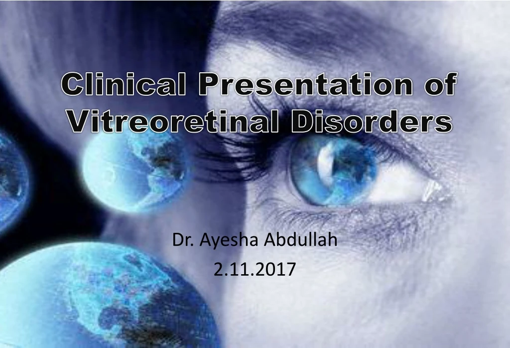
Clinical Presentation of Vitreoretinal Disorders Dr. Ayesha Abdullah 2.11.2017
Learning outcomes By the end of this lecture the students would be able to; • Identify the common symptoms and signs of VR disorders (VRD) and correlate them with the underlying problems with the structure and function of the VR. • Identify structural landmarks on retinal photographs. • Correlate the indications for commonly used investigations for the assessment of retinal disorders with the underlying pathology.
Common Presenting Symptoms in VRD • Visual loss, mostly painless, sudden/ gradual • Loss of central vision • Loss of peripheral vision • Loss of visual field • Loss of colour vision • Distorted vision; metamorphopsia, micropsia, macropsia
Common Presenting Symptoms in VRD • Loss of contrast sensitivity • Glare sensitivity • Night blindness • Photopsia/ flashes • Floaters ; 'specks', 'flies', 'spiders' and, ‘cobweb‘, ‘mosquitoes’.
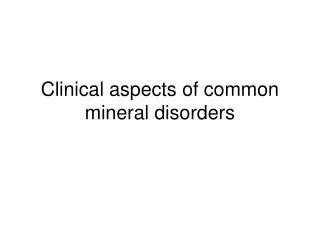
Loss of central vision http://www.retina-international.org/
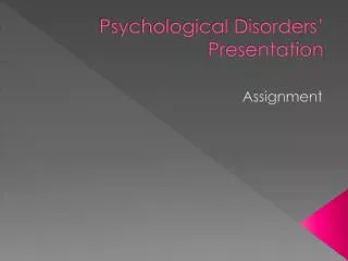
Loss of peripheral vision http://www.retina-international.org/
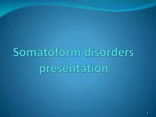
Colour vision deficiency http://www.achromatopsia.info/childrens-vision/
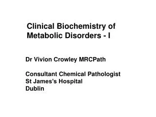
Scotoma Negative Scotoma Positive Scotoma
Commonly used investigations B Scan A Scan