Download

1 / 14
150 likes | 263 Views
Autoantibody Signature in Human Ductal Pancreatic Adenocarcinoma. Wangzhen 07.12.05. 1. Introduction.

E N D
Autoantibody Signature in Human Ductal PancreaticAdenocarcinoma Wangzhen 07.12.05
1. Introduction • Pancreatic ductal adenocarcinoma (PDAC) is a highly aggressive malignancy characterized by rapid progression, invasiveness, and resistance to treatment. Due to its early and asymptomatic invasion and metastasis, 80% of patients are inoperable at diagnosis and die within a few months. • It is impossible to diagnose PDAC at an early stage due to the lack of a reliable marker. There is thus a need for better early detection systems. • This study was designed to use a proteomics-based approach as a means of identifying antigens that elicit a humoral response in PDAC patients and search for suitable targets for early PDAC diagnosis and immunotherapy.
2. Methods 肿瘤血清蛋白质组分析法 (Tumor serologic proteome analysis, SERPA) • 一种将肿瘤免疫学与蛋白质组学相结合的方法 • 首先采用双向凝胶电泳(2-DE)分离肿瘤组织、癌旁正常组织的蛋白质,2-DE胶转膜后与患者或正常人血清中的某类免疫球蛋白建立Western blot反应图谱,通过计算机分析确定差异反应的蛋白质斑点,然后采用质谱分析和生物信息学方法对平行胶中相应的差异蛋白质点进行鉴定,筛选出肿瘤相关抗原。最后,用其他肿瘤组织和正常组织对肿瘤相关抗原的特异性进行分析。
3. Materials Serum samples: 70 PDAC patients, 40 healthy subjects(HS), 30 non-PDAC tumor patients , 15 chronic pancreatitis(CP) Cell lines: CF-PAC-1(metastatic), MiaPaCa-2(undifferentiated) and BxPC-3(poorly differentiated) were derived from PDAC patients Antibodies: mouse monoclonal anti-triosephosphateisomerase 1, mouse monoclonal anti-Keratin 10 antibody, rabbit polyclonal anti-cofilin-1 antibody HRP-conjugated rabbit anti-human IgG antibody, HRP-conjugated goat anti-mouse IgG antibody, HRP-conjugated goat anti-rabbit IgG antibody
4. Experimental Process Ⅰ. Sample preparation Ⅱ. 2-DE and WB with sera Ⅲ. Protein identification Ⅳ. Protein analysis
2-DE map images 2-DE WB images WB(1.serum 2. HRP-conjugated rabbit anti-human IgG antibody) 2-DE WB(1. specific antibodies 2. HRP-conjugated goat anti-mouse IgG antibody or HRP-conjugated goat anti-rabbit IgG antibody) MS WB analysis of PDAC sera reactivity against recombinant COF1 WB and immunohistochemical analysis of protein expression in tumor and normal pancreatic tissues Protein lysate
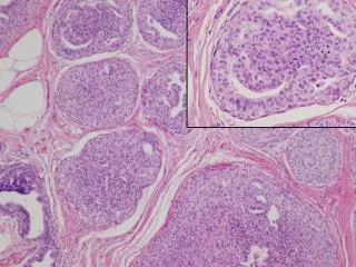
5. Results 5.1 Sera from PDAC patients contain autoantibodies to tumor proteins Figure 1. 2-DE map and Western blot of CF-PAC-1 and MiaPaCa-2 cell lines. The proteins from CF-PAC-1 cell line were separated by 2-DE and silver stained (A) or transferred to a nitrocellulose membrane and probed with PDAC patient (B and C) or HS (D) sera. The proteins from MiaPaCa-2 cell line were separated by 2-DE and silver stained (E) or transferred to a nitrocellulose membrane and probed with PDAC patient sera (F, G, and H).
Figure 2. Validation of PDAC serum reactivity against COF1, TPIS, and K1C10 by WB analysis. The reactivity against COF1 by PDAC sera (N ) 19) was tested on recombinant GST-COF1 by 1-D WB. (A) Representative result of 1-D WB with anti-cofilin-1 antibody, line 1, and with a PDAC serum, line 2. (B) Cropped images from CF-PAC-1 2-DE and 2-DE WB with anti-TPIS, anti-K1C10, and anti-COF1 antibody.
5.2 Expession of identified proteins in tumor and normal pancreatic tissues
6. Discussion A serological approach that combined 2-DE expression profiling of three human pancreatic tumor cell lines (CFPAC-1, MiaPaCa-2, and BxPC-3) and WB with PDAC patient sera was used to look for autoantibodies to PDAC-associated antigens. The study found that from 11 to 27% of PDAC patient sera contain IgG against two functional kinds of proteins: metabolic enzymes (TPIS, AL1A1, G6PD, IDCH, and EFTU) and cytoskeletal proteins (K1C10 and COF1).
In our study, antibodies to the identified proteins were not detected in sera from HS and non-PDAC tumor patients, suggesting that the antibody response to these proteins is characteristic of PDAC. As the most frequent antibody response in PDAC patients is directed to COF1 and TPIS2, our data support the hypothesis that the two proteins play a role in the biology of pancreatic cancer.