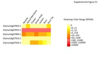
Download
1 / 21
220 likes | 447 Views
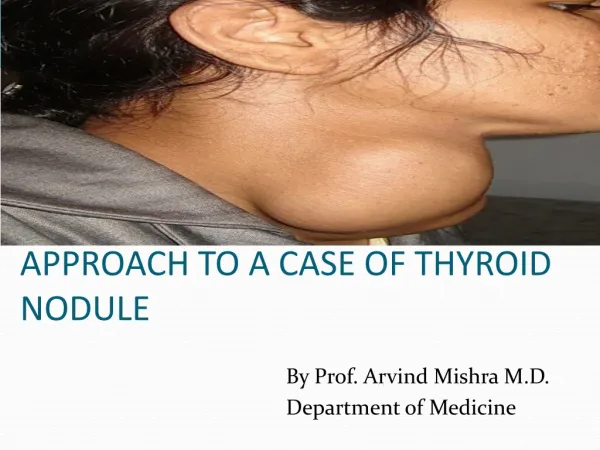
Approach to a thyroid nodule. Andy Sher PGY-2 Family Medicine. Case. 44 y.o. woman, 2 cm nodule palpable in left lobe of thyroid gland at annual exam – smooth, non-tender. No lymphadenopathy No symptoms of hyper/hypo thyroid. No compressive symptoms Past Med Hx: HTN Meds: HCTZ

E N D
Approach to a thyroid nodule Andy Sher PGY-2 Family Medicine
Case • 44 y.o. woman, 2 cm nodule palpable in left lobe of thyroid gland at annual exam – smooth, non-tender. No lymphadenopathy • No symptoms of hyper/hypo thyroid. No compressive symptoms • Past Med Hx: HTN • Meds: HCTZ • Fam Hx: no hx of thyroid disease
Epidemiology • Palpable thyroid nodules – 4-7% of population • Prevalence 19-67% - based on nodules found incidentally on ultrasound • 4:1 women:men
Epidemiology • Geographic areas with iodine deficiency • Thyroid carcinoma in 5-10% of palpable nodules • Following ionizing radiation, nodules develop at a rate of 2% annually
Presentation • Majority are asymptomatic • <1% cause hyperthyroidism • Neck pressure or pain if spontaneous hemorrhage
History • Symptoms of hyper or hypothyroidism • Previous nodules, goiters, family history of autoimmune thyroid disease, thyroid carcinoma, or familial polyposis • Hashimoto’s thyroiditis – association with thyroid lymphoma
History – Red Flags • Male • < 20 years, > 65 years • Rapid growth of nodule • Symptoms of local invasion (dysphagia, neck pain, hoarseness) • Hx of radiation to head or neck • Family hx of thyroid CA or polyposis
Physical Exam • Less than 1 cm usually not palpable • ½ of all nodules detected by ultrasonography not detected by physical exam • Should also examine for lymphadenopathy
Physical Exam • Smooth or nodular • Diffuse or localized • Soft or hard • Mobile or fixed • Painful or non-tender
Laboratory • TSH • Serum calcitonin if family hx of medullary thyroid carcinoma • Do not use thyroid function tests to differentiate benign from malignant
Radiology • Ultrasound • to document size, location, and character of nodule • To determine changes in size of nodules over time or to detect recurrent lesions • U/S guided biopsy decreases the incidence of indeterminate specimens
Radiology • Thyroid scan • Can not reliably distinguish benign from malignant nodules • Cold nodules – 5-15% are malignant • Hot nodules – almost always benign
Fine Needle Aspiration • Should be 1st test in the euthyroid patient • Sensitivity 68-98% • Specificity 72-100% • False negative rate 1-11% • False positive rate 1-8% • Sampling errors in very large and very small nodules – minimized by u/s guided biopsy
Treatment • Surgical treatment indications • Malignancy • Indeterminate cytology and suspicious H&P • Indeterminate cytology and “cold nodule” • Toxic nodules (suppression of TSH, symptoms – a-fib) – can use radioactive iodine or surgery • Repeated recurrence of cystic lesions
Treatment • Benign biopsies – can be followed without surgery and monitored q 6 months by physical exam, u/s • Surveillance – change in nodule size and symptoms – repeat FNA if nodule grows.
Suppression treatment • Post-operative suppression treatment following resection of cancer • TSH should be maintained for target of 0.5 mU per L • Greater suppression for high risk patients, metastatic or locally invasive not completely removed
Suppression treatment • For benign solitary nodule controversial • Follow at 6 month intervals • Thyroxine to suppress TSH to 0.1 to 0.5 mU per L for 6-12 months • After 12 months, maintain TSH in low normal range
Incidental Nodule on U/S • Most are benign and can be monitored without further testing • FNA if • nodule becomes palpable • findings suggestive of malignancy on u/s • larger than 1.5 cm • Hx of head or neck irradiation • Strong family hx of thyroid cancer
Case • 44 y.o. woman, 2 cm nodule palpable in left lobe of thyroid gland at annual exam – smooth, non-tender. No lymphadenopathy • TSH ordered – normal • Thyroid u/s – confirms 2 cm nodule, solid • FNA - benign
Case • Repeat U/S at 1 year – nodule now 2.5 cm in size • Repeat FNA – benign • Could consider suppression therapy, or continue to follow.