Download
1 / 85
850 likes | 1.06k Views
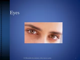
Head and Eyes. Dr. Megan Gonzales ND, EAMP SIOM Western Clinical Sciences Fall 2012. External Eye Anatomy. Internal Eye Anatomy. Eyelid and Lacrimal Ducts. Blepharitis Exopthalmos Ptosis Orbital Cellulitis Dacrocystitis Chalazion Stye. Blepharitis.

E N D
Head and Eyes • Dr. Megan Gonzales ND, EAMP • SIOM Western Clinical Sciences Fall 2012
Eyelid and Lacrimal Ducts • Blepharitis • Exopthalmos • Ptosis • Orbital Cellulitis • Dacrocystitis • Chalazion • Stye
Blepharitis is from an overgrowth of bacteria and may be linked to repeated styes and chalazia. You are more likely to develop this condition if you have seborrheic dermatitis of the face or scalp, rosacea, lice, and allergies.
Exophthalmos • Exophthalmos (also called exophthalmia or proptosis) is a bulging of the eye anteriorly out of the orbit. Exophthalmos can be either bilateral (as is often seen in Graves' disease) or unilateral (as is often seen in an orbital tumor).
other causes - infection, parasitic cysts, pseudoexophthalmos related to paralysis of the extraocular muscles • Signs and symptoms: bulging eyeballs, with diplopia, extraocular muscle edema causes malallignment of the eyes. Other symptoms are related to systemic cause of exophthalmos (hyperthyroidism, tumor, cavernous sinus thrombosis) • Diagnosis: usually based on obvious sign of bulging eyes, CT or MRI, culture of any discharge, Bx of orbital tissue • Treatment: depends on underlying cause of dysfunction. Cold compress for trauma, treatment with antimicrobials, surgery, partial or total thyroidectomy
Ptosis • PTOSIS: is also called "drooping eyelid." It is caused by weakness of the muscle responsible for raising the eyelid, damage to the nerves that control those muscles, or looseness of the skin of the upper eyelids.
can be congenital either genetic or through a trait where the levator palpebrae muscles fail to fully develop • aquired: advanced age - from cataract surgery, eyelid edema or other mechanisms leading to a heavy eyelid • muscular dystrophy or myasthenia gravis where muscles are malfunctional • paralysis • nutritional factors such as thiamine deficiency in chronic alcoholism and other states of malnutrition • can lead to lazy eye in children • diagnosis is based on physical exam and finding the underlying condition causing ptosis • may not require treatment but will be surgically correct if it interferes with vision. Can also be prescribed special glasses that contain and eyelid “crutch”
Orbital Cellulitis • Orbital cellulitis is an acute infection of the tissues immediately surrounding the eye, including the eyelids, eyebrow, and cheek.
related to any type of infection from direct inoculation via the blood stream or near-by structures eg. sinuses. Also surgery, insect or animal bites, and foreign body trauma • can cause cavernous sinus thromosis, hearing loss, septicemia, meningitis, optic nerve damage • usually unilateral eyelid edema, reddened eyelids, hyperemic orbital tissue, extreme orbital pain, impaired eye movement, purulent discharge. Associated: fever, chills, malaise according to the cause of cellulitis. • Diagnosis is based on clinical picture, CT and MRI of sinuses and brain, culture and sensitivity of discharge or wounds. • Treatment: antimicrobials both systemic and topical, moist compresses, bed rest and fluids, I & D if abscess is present
Dacryocystitis is an infection of the nasolacrimal sac, frequently caused by nasolacrimal duct obstruction. The term derives from the Greek dákryon (tear),[1] cyst (sac), and -itis (inflammation). It causes pain, redness, and swelling over the inner aspect of the lower eyelid. It is most commonly caused by Staphylococcus aureus and Streptococcus pneumoniae. The most common complication is corneal ulceration, frequently in association with S. pneumoniae.
can lead to orbital cellulitis • it is extremely painful and presents with constant tearing, there can be a sensation of pain or pressure over the nasolacrimal sac and applied pressure may or may not cause discharge from the punctum • clinical picture, CBC and culture and sensitivity of any discharge is used to determine type of infection, in infants if it is due to duct atresia and x-ray after radiopaque injection shows the location • treatment: warm compresses with topical and systemic antimicrobials, surgery can also be performed.
A chalazion (a more intense stye) is a small lump in the eyelid caused by obstruction of an oil producing or meibomian gland. Chalazia may occur in the upper or lower lids, causing redness, swelling and soreness in some cases.
can lead to astigmatism • diagnosis: visual exam and palpation of the eyelid. A Bx should be performed if the chalazion is persistent or recurring to rule out meibomian cancer. • Treatment: warm compresses for 10-15 minutes up to 4 times a day. Incision and curettage may be necessary. After surgery antibiotic topicals may be prescribed but otherwise are not useful.
Hordeolum AKA Stye • Stye (hordeolum) A stye is caused by bacteria from the skin that get into the oil glands in the eyelids that provide lubrication to the tear film. Styes are similar to common acne pimples that occur elsewhere on the skin. You may have more than one stye at the same time. • Styes usually develop over a few days and may drain and heal on their own. A stye can become a chalazion -- this is when an inflamed oil gland becomes fully blocked. If a chalazion gets large enough, it can cause trouble with your vision.
Conjunctival Disorders • inclusion conjunctivitis • conjunctivitis • trachoma
Inclusion Conjunctivitis • caused by Chlamydia trachomatis an obligate intracellular organism, can be a cause of “ophthalmia neonatorum” due to infection while passing through the birth canal. • usually occurs in adults 18-30 years of age • can cause otitis media and blindness. • in neonates the first signs are reddened eyelids and slight discharge. A psuedomembrane can form which causes conjunctival scarring. • In adults follicles appear inside the lower eyelids. It can persist for weeks or months with superficial corneal involvement.
Inclusion Conjunctivitis • Diagnosis is based on clinical features and history of sexual behavior including contact with an infected individual, a conjunctival scraping will be performed to check for the specific organism. • A systemic anti-microbial is prescribed generally erythromycin and neonates are treated with anti-microbial ointment to the eyelids at 1 hour post birth.
Conjunctivitis, commonly known as pink eye, is an infection of the conjunctiva (the outer-most layer of the eye that covers the sclera). The three most common types of conjunctivitis are: viral, allergic, and bacterial. Each requires different treatments. With the exception of the allergic type, conjunctivitis is typically contagious and self-limiting. • Common bacterial causes: Staph aureus, Neisseria gonorrhoeae • Common viral causes: adenoviruses, herpes simplex type 1
complications include: corneal infiltrates, reinfection and eye loss • hyperemia of the conjunctiva with tearing and sometimes discharge, pain and photophobia. It often begins in one eye but spreads quickly to the other. Acute bacterial conjunctivitis lasts about 2 weeks, viral conjunctivitis has little to no exudate and can produce a severe disabling disease or be 2-3 weeks in course. • Diagnosis -symptoms, stained conjunctival scrapings revealing lymphocytes due to viruses, neutrophils with a bacterial infection and eosinophils when allergens are the culprit. • Treatment: application of broad-spectrum anti-biotics, prevention of a secondary infection with viral infections. The most important thing is to prevent spread of the disease. • As Neisseria gonorrhoeae can be an infective agent diagnosis is important and the health care provider must report the infection!
Trachoma • the most common cause of preventable blindness in less developed nations • a form of chronic keratoconjunctivitis, may have a systemic symptomatic picture • it is self limiting but permanently damages the cornea and conjunctiva due to scarring, the resulting secondary infections can cause blindness it is therefore important to diagnose and treat the condition early • this is a result of an infection from Chlamydia trachomatis - gram negative obligate intracellular bacterium. Transmitted eye-to-eye by flies and gnats or hand-to-eye contact.
it is prevalent in Africa, Latin America and Asia primarily in children. • Can lead to conjunctival scarring and corneal scarring, eyelid deformities and loss of vision • it begins with a mild-looking bacterial conjunctivitis. After 1 month untreated the conjunctival follicles become enlarged and yellow and gray. At this time small blood vessels invade the cornea and upper lid. • Contracture of the eyelid leads to entropion (the eyelids turn in and eyelashes scratch the cornea) • Diagnosis is made based on symptoms and signs confirmed with a special stain detecting chlamydia • Treatment requires topical and systemic antibiotic medications with erythromycin, doxycycline or sulfonamides and surgery for the entropion.
Corneal Disorders • Keratitis • Corneal Abrasion • Corneal ulcer
Keratitis • inflammation of the cornea it may result from bacterial, fungal or viral infections and can lead to blindness • commonly caused by the herpes simplex virus type 1. Bacterial infections can result due to infection of a corneal abrasion. • can cause blindness and corneal scarring and perforation • it is usually unilateral, presenting with decreased vision and discomfort to acute pain and tearing with photophobia. • the corneal light reflexes may be distorted when examined with a penlight.
it is diagnosed by patient history - a recent URI with cold sores and eye irritation after wearing contacts. • Treatment - if due to herpes antiviral drops , ointment or oral acyclovir is prescribed. Bacterial infections require antibacterial drops given every half hour for the first 48 hours.
Corneal Abrasions • a scratch on the epithelium of the cornea, usually due to foreign body under the eyelid. They happen in people who don’t wear protective eye coverings in high risk fields or when people fall asleep with contacts in. Can also be due to accidental dust, dirt or grit. The most common cause of ophthalmologic EMERGENT hospital visits in the US. • Can cause corneal erosion, ulceration or permanent vision loss
causes erythema, increased tearing, discomfort on blinking, feeling of “something in the eye”, pain disproportionate to the size of injury secondary to high innervation from the trigeminal nerve. • Diagnosis is made based on history of eye trauma or prolonged use of contacts and symptoms of an abrasion. An ophthamologist will use a stain and special light to discern scratches. • Treatment: topical anesthetics, removal of foreign body, antibiotic eye drops, a pressure patch to relieve pain on blinking, discontinued use of contacts while healing.
Corneal Ulcers • a major cause of blindness worldwide. They produce scarring or perforation. Ulcers may occur anywhere but are most commonly found marginally. They require treatment within hours to prevent visual impairment. • Usually due to a variety of infective agents. Commonly: Staph aureus, Pseudomonas aeruginosa, Herpes simplex type 1, varicella-zoster, Candida and Cephalosporium (bacterial, viral and fungal). • Can cause corneal scarring, loss of the eye, loss of vision
begins with pain that is worse on blinking, photophobia and increased tearing. Eventually a central ulceration will cause blurred vision. The eye may be injected and purulent discharge may be present. • Diagnosis is based on history of trauma or over use of contact lenses. Use of a dye to show the ulcer confirms diagnosis. • Treatment: PROMPT is NECESSARY TO SAVE VISION. Broad spectrum antimicrobial drops are used. If due to bacterial infection a patch SHOULD NOT be used because it generates an even better growth environment. Analgesics and observation for possible development of secondary glaucoma.
Uveal tract, Retinal, Lens Disorders • uveitis • retinal detachment • vascular retinopathies • age-related macular degeneraton • cataract • retinitis pigmentosa
Uveitis is swelling and irritation of the uvea, the middle layer of the eye. The uvea provides most of the blood supply to the retina.
Uveitis • usually idiopathic but can be the result of allergies, bacteria, viruses, fungi, chemicals, trauma and even surgery. • can cause cataracts, glaucoma, retinal detachment, blindness • moderate to severe unilateral eye pain, severe injection, tearing, small pupil non-reactive to light, blurred vision and sometimes produces deposits left on the back of the cornea. Onset is acute or insidious. • Diagnosis is made with slit lamp examination. • Treatment: is vigorous and prompt. One must ascertain the cause. For severe cases oral corticosteroids are prescribed.