Download
1 / 40
400 likes | 513 Views
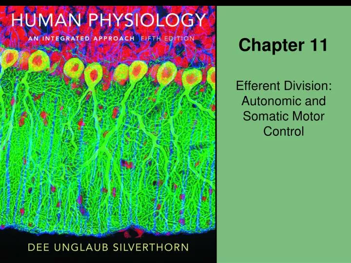
Chapter 11. Efferent Division: Autonomic and Somatic Motor Control. About this Chapter. Autonomic division Autonomic reflexes Antagonistic controls Control of cardiac and smooth muscle, and glands in homeostasis Agonists and antagonists in research and medicine Somatic motor division

E N D
Chapter 11 Efferent Division: Autonomic and Somatic Motor Control
About this Chapter • Autonomic division • Autonomic reflexes • Antagonistic controls • Control of cardiac and smooth muscle, and glands in homeostasis • Agonists and antagonists in research and medicine • Somatic motor division • CNS control of skeletal muscles through neuromuscular junctions
Role of the Autonomic Division in Homeostasis • Antagonistic branches • Parasympathetic • “Rest and digest” • Restore body function • Sympathetic • “Fight or flight” • Energetic action
Role of the Autonomic Division in Homeostasis Figure 11-1
The Hypothalamus, Pons, and Medulla Initiate Autonomic, Endocrine, and Behavioral Responses • Coordination of homeostatic responses • Autonomic • Endocrine • Behavioral Figure 11-2
Autonomic Control Centers in the Brain • Hypothalamus • Water balance, temperature, and hunger • Pons • Respiration • Medulla • Respiration • Cardiac • Vomiting • Swallowing Temperature control Water balance Eating behavior Hypothalamus Pons Urinary bladder control Medulla Secondary respiratory center Blood pressure control Respiratory center Figure 11-3
Autonomic Pathways Figure 11-4
Antagonistic Control of the Autonomic Division • Most internal organs are under antagonistic control • One autonomic branch is excitatory and the other branch is inhibitory • Example: • Effector organ: heart • Parasympathetic response: slows rate • Sympathetic response: increases rate and force of contraction
Autonomic Sympathetic and Parasympathetic Pathways Hypothalamus, Reticular formation Ganglion Pons Medulla Vagus nerve Spinal cord Sympathetic chain Pelvic nerves KEY Parasympathetic Sympathetic Figure 11-5
Autonomic Sympathetic and Parasympathetic Pathways • Sympathetic versus parasympathetic pathways • Spinal cord exit • Neurotransmitters • Receptors • The major parasympathetic tract is the vagus nerve Medulla Vagus nerve Left lung Right lung Liver Spleen Stomach Pancreas Proximal two-thirds of colon Entire small intestine Figure 11-6
Autonomic Sympathetic and Parasympathetic Pathways Sympathetic pathways use acetylcholine and norepinephrine. Parasympathetic pathways use acetylcholine. CNS ACh Nicotinic receptor Autonomic ganglion Norepinephrine ACh Adrenergic receptor Muscarinic receptor T T Target tissue Figure 11-7
Sympathetic and Parasympathetic Neurotransmitters Table 11-1
Autonomic Targets • Autonomic pathways control: • Smooth muscle • Cardiac muscle • Exocrine glands (select) • Endocrine glands (select) • Lymphoid tissue • Adipose tissue
Autonomic Neuron Structure • Neuroeffector junction • Postganglionic axon • Varicosities • Axon • Neurotransmitter synthesis
Varicosities in Autonomic Neurons Axon of postganglionic autonomic neuron Vesicle containing neurotransmitter Mitochondrion Varicosity Figure 11-8 Varicosities Smooth muscle cells Figure 11-8
Norepinephrine Release at a Varicosity of a Sympathetic Neuron 1 Action potential arrives at the varicosity. 2 Depolarization opens voltage-gated Ca2+ channels. Axon varicosity MAO 3 Ca2+ entry triggers exocytosis of synaptic vesicles. Tyrosine 8 7 Axon Voltage-gated Ca2+ channel NE 1 Action potential 4 NE binds to adrenergic receptor on target. 3 Exocytosis Active transport 5 Receptor activation ceases when NE diffuses away from the synapse. Ca2+ 2 6 NE 5 Diffuses away 4 Blood vessel NE is removed from the synapse. 6 G Response Target cell Adrenergic receptor 7 NE can be taken back into synaptic vesicles for re-release. 8 NE is metabolized by monoamine oxidase (MAO). Figure 11-9, steps 1–8
Sympathetic Branch: Stimulation • Pupil dilation • Salivation • Heart beat and volume • Blood vessel and bronchiole dilation • Fat breakdown • Ejaculation
Sympathetic Branch: Inhibition • Digestion • Pancreas secretion • Urination
Adrenal Medulla • Primary neurohormone • Epinephrine • Multiple and distant targets
The Adrenal Medulla Adrenal cortex is a true endocrine gland. Adrenal medulla is a modified sympathetic ganglion. Adrenal gland Kidney (b) (a) The chromaffin cell is a modified postganglionic sympathetic neuron. ACh Blood vessel Preganglionic sympathetic neuron Spinal cord Epinephrine is a neurohormone that enters the blood. To target tissues (c) Adrenal medulla Figure 11-10
The Adrenal Medulla Adrenal gland Kidney (a) Figure 11-10a
The Adrenal Medulla Adrenal cortex is a true endocrine gland. Adrenal medulla is a modified sympathetic ganglion. (b) Figure 11-10b
The Adrenal Medulla The chromaffin cell is a modified postganglionic sympathetic neuron. ACh Blood vessel Preganglionic sympathetic neuron Spinal cord To target tissues Epinephrine is a neurohormone that enters the blood. (c) Adrenal medulla Figure 11-10c
Parasympathetic Branch • Acetylcholine • Muscarinic receptors • G protein-coupled • Second messenger pathways • At least five subtypes
Parasympathetic Branch: Actions • Constricts pupils and bronchioles • Slows heart • Stimulates • Digestion • Insulin release • Urination • Erections
Autonomic Agonists and Antagonists • Agonists and antagonists are important tools in research and medicine Table 11-3
Efferent Pathways of the Peripheral Nervous System SOMATIC MOTOR PATHWAY AUTONOMIC PATHWAYS Parasympathetic pathway Sympathetic pathways Adrenal sympathetic pathway CNS CNS CNS CNS ACh Adrenal cortex Nicotinic receptor Adrenal medulla Ganglia E ACh Nicotinic receptor Ganglion receptor NE ACh Muscarinic receptor Autonomic effectors: Blood vessel • Smooth and cardiac muscles • Some endocrine and exocrineglands • Some adipose tissue 1 receptor E ACh 2 receptor Nicotinic receptor KEY ACh= acetylcholine E= epinephrine NE= norepinephrine Skeletal muscle Figure 11-11
Efferent Pathways of the Peripheral Nervous System AUTONOMIC PATHWAYS Parasympathetic pathway Sympathetic pathways Adrenal sympathetic pathway CNS CNS CNS ACh Adrenal cortex Nicotinic receptor Adrenal medulla Ganglia E ACh Nicotinic receptor Ganglion receptor NE ACh Muscarinic receptor Autonomic effectors: Blood vessel • Smooth and cardiac muscles • Some endocrine and exocrineglands • Some adipose tissue 1 receptor E 2 receptor KEY ACh= acetylcholine E= epinephrine NE= norepinephrine Figure 11-11 (2 of 5)
Efferent Pathways of the Peripheral Nervous System Figure 11-11 (3 of 5)
Efferent Pathways of the Peripheral Nervous System Figure 11-11 (4 of 5)
Efferent Pathways of the Peripheral Nervous System Figure 11-11 (5 of 5)
Somatic versus Autonomic Divisions Table 11-5
Somatic Motor Division • Single neuron • CNS origin • Myelinated • Terminus • Branches • Neuromuscular junction
Somatic Motor Division Figure 11-11 (1 of 5)
Anatomy of the Neuromuscular Junction Somatic motor neuron The neuromuscular junction Muscle fiber Terminal bouton Figure 11-12 (1 of 3)
Anatomy of the Neuromuscular Junction Schwann cell sheath Axon terminal Mitochondria Motor end plate Figure 11-12 (2 of 3)
Anatomy of the Neuromuscular Junction Synaptic vesicle (ACh) Presynaptic membrane Synaptic cleft Postsynaptic membrane Nicotinic ACh receptors Figure 11-12 (3 of 3)
Events at the Neuromuscular Junction Somatic motor neuron Axon terminal Ca2+ Ca2+ Action potential ACh Acetyl + choline Voltage-gated Ca2+ channel Skeletal muscle fiber AChE Motor end plate Nicotinic receptor (a) Figure 11-13a
Events at the Neuromuscular Junction Open channel Closed channel K+ Na+ ACh K+ Na+ (b) Figure 11-13b
Cigarette Smoking Among American High School Students Questions 11-1