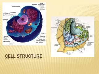
Cell Structure
Cell Structure. Components structure and function. Endoplasmic Reticulum. Smooth and Rough ER. Endoplasmic Reticulum. Extensive membrane system which runs through the cytoplasm Much of the ER has ribosomes attached to it which is called Rough ER Smooth ER do not have ribosomes attached

Cell Structure
E N D
Presentation Transcript
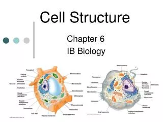
Cell Structure Components structure and function
Endoplasmic Reticulum Smooth and Rough ER
Endoplasmic Reticulum • Extensive membrane system which runs through the cytoplasm • Much of the ER has ribosomes attached to it which is called Rough ER • Smooth ER do not have ribosomes attached • SER and RER have different functions • RER – transports proteins made by ribosomes • SER – makes lipids and steroids (cholesterol & reproductive hormones) • The membranes form a system of flattened sacs, like sheets called cisternae • The space inside the sacs forms a compartment separate from the surrounding cytoplasm
ER cont’d • ER is continuous with the nuclear envelope • The cisternae goes on to form the Golgi apparatus • RER tends to be sheet-like • SER tends to be tubular • SER has different roles in different cells • RER tends to be extensive in cells which produce a lot of protein (enzyme-secreting cells in the digestive system) • RER gives more surface area for protein synthesis, storage (Ca) and transportation
Ribosomes Sites of Protein Synthesis
Ribosomes • There are 2 types • 70S (found in prokaryotes, chloroplasts and mitochondria) • 80S (found in eukaryotes) • They are very small organelles • Consist of 2 units (see BS P. 150) • Made of RNA (ribonucleic acid) and protein • May form polysomes which is a collection of ribosomes strung along mRNA
Ribosomes cont’d • Sites for protein synthesis • Found free in the cytoplasm as well as on the RER. • Proteins that are manufactured on the ribosomes are transported throughout the cell by RER
Ribosomes cont’d • Proteins made by ER bound ribosomes tend to be secreted from the cell. • Haemoglobin is an example of a protein made by free ribosomes in the cytoplasm in young RBC • Enzymes involved in glycolysis (1st stage in respiration)
Quick Question! • List 4 functions of ER • To form an extensive transport system throughout the cell (connecting nuclear envelope to cell membrane structure • Production and packaging of proteins (RER) • Synthesis of lipids and steroids (SER) • Collection, storage and distribution of these materials
Mitochondrion Powerhouse of the Cell
Mitochondria • Surrounded by 2 membranes (an envelope) • The inner forms the cristae (finger like projections) • The interior is filled with a fluid or matrix • Main function: • To carry out the later stages of aerobic respiration (make ATP) • Another function is to make lipids • Contained in cells which require a lot of energy • The membrane of the mitochondria is the site of ATP synthesis • The matrix of the mitochondria is the site for the Kreb’s cycle
Mitochondria cont’d • The number of mitochondria found in cells depends on the type of organism and the nature of the cell • Their shapes and sizes vary from • Spiral • Spherical • Elongated • Cup-shaped • Branched • Length range from 1.5-10 micrometer but diameter does not exceed 1 micrometer
Mitochondria cont’d • Contains 70S ribosomes • Contains circular DNA • Able to change shape • Able to move to areas in the cell where a lot of activity is taking place • Some can remain fixed (insect flight muscle) • See BS p. 276 for further details on structure of mitochondria
Golgi Body/Apparatus • A series of flattened membrane vesicles • Formed from the endoplasmic reticulum • Transports proteins from the RER to the cell membrane for export • Part of the RER containing proteins fuse with one side of the Golgi body membranes • The point at which fusion occurs their contents are released via exocytosis
Chloroplast • Bigger and fatter than mitochondria • Site of photosynthesis • Found in photosynthetic organisms (plant & algae) • Enclosed in a double membrane • Thylakoid membranes are folded into thylakoid disks • Stacked into piles of grana • The space between the inner membrane and the thylakoid is called the stroma • Thylakoid membranes contain chlorophyll and stalk elements
Chloroplasts cont’d • Thylakoids are the site of photosynthesis and ATP synthesis • Chloroplasts also contain • starch grains • Ribosomes • Circular DNA
Lysosomes • Small membrane-bound vesicles • Formed from RER containing a cocktail of digestive enzymes • Break down unwanted chemicals, toxins, organelles or even whole cells - recycling materials • Can fuse with a feeding vacuole to digest its contents
Cell membrane • Also called plasma membrane or cell surface membrane • Thin & flexible • Found around the outside of all cells • Made of phospholipids, proteins and carbohydrates arranges in a fluid mosaic structure • Separates the contents of the cell from the outside environment • Controls the entry and exit of materials
Cell membrane cont’d • Responsible for many properties of the cell • Membranes that surround the nucleus and other organelle are almost identical to the cell membrane
Phospholipids • The phospholipids are arranged in a bilayer, • polar, hydrophilic phosphate heads facing outwards, • non-polar, hydrophobic fatty acid tails facing each other in the middle of the bilayer.
This hydrophobic layer acts as a barrier to all but the smallest molecules • effectively isolating the two sides of the membrane. • Different kinds of membranes can contain phospholipids with different fatty acids, affecting the • strength and • flexibility of the membrane, • Animal cell membranes also contain cholesterol linking the fatty acids together and so stabilising and strengthening the membrane.
The Proteins • usually span from one side of the phospholipid bilayer to the other (intrinsic proteins), • but can also sit on one of the surfaces (extrinsic proteins). • They can slide around the membrane very quickly and collide with each other, but can never flip from one side to the other. • have hydrophilic amino acids in contact with the water on the outside of membranes, and hydrophobic amino acids in contact with the fatty chains inside the membrane. • comprise about 50% of the mass of membranes, and are responsible for most of the membrane's properties.
Proteins cont’d • Proteins that span the membrane are usually involved in transporting substances across the membrane • Proteins on the inside surface of cell membranes are often attached to the cytoskeleton and are involved in maintaining the cell's shape, or in cell motility. • They may also be enzymes, catalysing reactions in the cytoplasm.
Proteins cont’d • Proteins on the outside surface of cell membranes can act as receptors by having a specific binding site where hormones or other chemicals can bind. • This binding then triggers other events in the cell. • They may also be involved in cell signalling and cell recognition, or • they may be enzymes, such as maltase in the small intestine (more in digestion).
Carbohydrates • found on the outer surface of all eukaryotic cell membranes, • are usually attached to the membrane proteins. • Proteins with carbohydrates attached are called glycoproteins. • The carbohydrates are short polysaccharides composed of a variety of different monosaccharides, and form a cell coat or glycocalyx outside the cell membrane. • The glycocalyx is involved in protection and cell recognition, and antigens such as the ABO antigens on blood cells are usually cell-surface glycoproteins.
Nucleus • This is the largest organelle. • Surrounded by a nuclear envelope, which is a double membrane with nuclear pores - large holes containing proteins that control the exit of substances such as RNA from the nucleus.
Nucleus cont’d • The interior is called the nucleoplasm, which is full of chromatin- a DNA/protein complex containing the genes. • During cell division the chromatin becomes condensed into discrete observable chromosomes. • The nucleolus is a dark region of chromatin, involved in making ribosomes.
Cytoskeleton • This is a network of protein fibres extending throughout all eukaryotic cells, used for support, transport and motility • The cytoskeleton is attached to the cell membrane and gives the cell its shape, as well as holding all the organelles in position • There are three types of protein fibres (microfilaments, intermediate filaments and microtubules), and each has a corresponding motor protein that can move along the fibre carrying a cargo such as organelles, chromosomes or other cytoskeleton fibres
Cytoskeleton • These motor proteins are responsible for such actions as: chromosome movement in mitosis, cytoplasm cleavage in cell division, cytoplasmic streaming in plant cells, cilia and flagella movements, cell crawling and even muscle contraction in animals.
Summary of the Differences Between Prokaryotic and Eukaryotic Cells
Endosymbiosis • Prokaryotic cells are far older and more diverse than eukaryotic cells. Prokaryotic cells have probably been around for 3.5 billion years - 2.5 billion years longer than eukaryotic cells. • It is thought that eukaryotic cell organelles like mitochondria and chloroplasts are derived from prokaryotic cells that became incorporated inside larger prokaryotic cells.
Endosymbiosis • This idea is called endosymbiosis, and is supported by these observations: • organelles contain circular DNA, like bacteria cells. • organelles contain 70S ribosomes, like bacteria cells. • organelles have double membranes, as though a single-membrane cell had been engulfed and surrounded by a larger cell.
Transport Across The Membrane • Cell membranes are a barrier to most substances, and this property allows materials to be concentrated inside cells, excluded from cells, or simply separated from the outside environment. • This is compartmentalization is essential for life, as it enables reactions to take place that would otherwise be impossible. Eukaryotic cells can also compartmentalize materials inside organelles.
Materials need to be able to enter and leave cells • There are five main methods by which substances can move across a cell membrane: • 1. Simple Diffusion • 2. Osmosis • 3. Facilitated Diffusion • 4. Active Transport • 5. Vesicles
A few substances can diffuse directly through the lipid bilayer part of the membrane. • The only substances that can do this are lipid-soluble molecules such as steroids, or very small molecules, such as H2O, O2 and CO2. • For these molecules the membrane is no barrier at all. • Since lipid diffusion is (obviously) a passive diffusion process, no energy is involved and substances can only move down their concentration gradient. • Lipid diffusion cannot be controlled by the cell, in the sense of being switched on or off.
Osmosis • Osmosis is the diffusion of water across a membrane. • It is in fact just normal lipid diffusion, but since water is so important and so abundant in cells (its concentration is about 50 M), the diffusion of water has its own name - osmosis. • The contents of cells are essentially solutions of numerous different solutes, and the more concentrated the solution, the more solute molecules there are in a given volume, so the fewer water molecules there are. • Water molecules can diffuse freely across a membrane, but always down their concentration gradient, so water therefore diffuses from a dilute to a concentrated solution.
Water Potential • Osmosis can be quantified using water potential, so we can calculate which way water will move, and how fast. • Water potential (Y, the Greek letter psi, pronounced "sy") is a measure of the water molecule potential for movement in a solution. • It is measured in units of pressure (Pa, or usually kPa), and the rule is that water always moves by osmosis from less negative to more negative water potential (in other words it's a bit like gravity potential or electrical potential). • 100% pure water has Y = 0, which is the highest possible water potential, so all solutions have Y < 0 (i.e. a negative number), and you cannot get Y > 0.
Cells and Osmosis • The concentration (or OP) of the solution that surrounds a cell will affect the state of the cell, due to osmosis. There are three possible concentrations of solution to consider: • Isotonic solution a solution of equal OP (or concentration) to a cell • Hypertonic solution a solution of higher OP (or concentration) than a cell • Hypotonic solution a solution of lower OP (or concentration) than a cell
The effects of these solutions on cells are shown in this diagram: