Download
1 / 66
690 likes | 715 Views
Learn about arteries, capillaries, and veins, their structure, types, and unique characteristics. Explore the roles of these blood vessels in transporting blood and exchanging molecules.

E N D
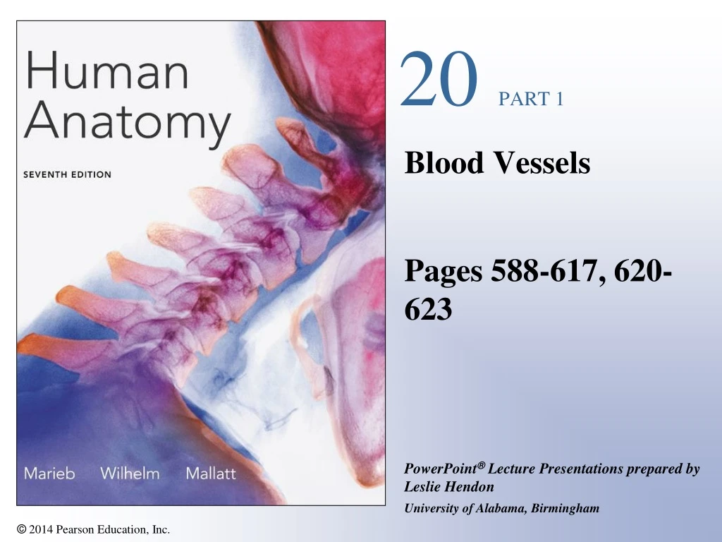
20 PART 1 Blood Vessels Pages 588-617, 620-623
Types of Blood Vessels • Arteries—carry blood away from the heart • Capillaries—smallest blood vessels • The site of exchange of molecules between blood and tissue fluid • Veins—carry blood toward the heart
Structure of Blood Vessels • Composed of three layers (tunics) • Tunica intima—composed of simple squamous epithelium • Tunica media—sheets of smooth muscle • Contraction—vasoconstriction • Relaxation—vasodilation • Tunica externa—composed of connective tissue • Lumen • Central blood-filled space of a vessel
Figure 20.1b Generalized structure of arteries, veins, and capillaries. Tunica intima Artery Vein Endothelium Subendothelial layer Internal elastic membrane Tunica media(smooth muscle andelastic fibers) External elastic membrane Tunica externa(collagen fibers) Vasa vasorum Valve Capillary network Lumen Lumen Basement membrane Capillary Endothelial cells
Figure 20.1a Generalized structure of arteries, veins, and capillaries. Artery Vein
Vasa vasorum Elastin Lumen Tunica externa Tunica media Tunica intima (a) Elastic artery (aorta, 12) Types of Arteries • Elastic arteries—the largest arteries • Diameters range from 2.5 cm to 1 cm • Includes the aorta and its major branches • Sometimes called conducting arteries • High elastin content dampens surge of blood pressure Figure 20.2a
External elastic membrane Internal elastic membrane Lumen Tunica externa Tunica media (b) Muscular artery (40) Types of Arteries • Muscular (distributing) arteries • Lie distal to elastic arteries • Diameters range from 1 cm to 0.3 mm • Includes most named arteries • Tunica media is thick • Unique feature • Internal and external elastic laminae Figure 20.2b
Types of Arteries • Arterioles • Smallest arteries • Diameters range from 0.3 mm to 10 µm • Larger arterioles possess all three tunics Lumen Endothelium Tunica media (c) Small arteriole (285) Figure 20.2c
Capillaries • Smallest blood vessels • Diameter from 8–10 µm • Red blood cells pass through single file • Site-specific functions of capillaries • Lungs—oxygen enters blood, carbon dioxide leaves • Small intestines—receive digested nutrients • Endocrine glands—pick up hormones • Kidneys—remove of nitrogenous wastes
Figure 20.1b Generalized structure of arteries, veins, and capillaries. Tunica intima Artery Vein Endothelium Subendothelial layer Internal elastic membrane Tunica media(smooth muscle andelastic fibers) External elastic membrane Tunica externa(collagen fibers) Vasa vasorum Valve Capillary network Lumen Lumen Basement membrane Capillary Endothelial cells
Figure 20.3 Red blood cells passing through a capillary (512).
Capillary Permeability • Endothelial cells—held together by tight junctions and desmosomes • Intercellular clefts—gaps of unjoined membrane • Small molecules can enter and exit • Two types of capillary • Continuous—most common • Fenestrated—have pores
Special Characteristics of Epithelia Narrow extracellular space Cilia Microvilli Apical region of an epithelial cell Cell junctions Tight junction Epithelium Adhesive belt Desmosome Gap junction Basal region Basal lamina Basement membrane Reticular fibers Nerve ending Connective tissue Capillary Figure 4.1
Pericyte Red blood cell in lumen Intercellular cleft Endothelial cell Basement membrane Tight junction Pinocytotic vesicles Endothelial nucleus (a) Continuous capillary. Least permeable and most common (e.g., skin, muscle). Structure of Capillaries—Cross Section Figure 20.4
Pinocytotic vesicles Red blood cell in lumen Fenestrations (pores) Endothelial nucleus Intercellular cleft Basement membrane Endothelial cell Tight junction (b) Fenestrated capillary. Large fenestrations (pores) increase permeability. Occurs in special locations (e.g., kidney, small intestine). Structure of Capillaries—Cross Section Figure 20.4
Sinusoids • Wide, leaky capillaries found in some organs • Usually fenestrated • Intercellular clefts are wide open • Occur in bone marrow and spleen • Sinusoids have a large diameter and twisted course
Endothelial cell Red blood cell in lumen Large intercellular cleft Tight junction Nucleus of endothelial cell Incomplete basement membrane (c) Sinusoidal capillary. Most permeable. Occurs in special locations (e.g., liver, bone marrow, spleen). Sinusoids Figure 20.4
Veins • Conduct blood from capillaries toward the heart • Blood pressure is much lower than in arteries • Smallest veins—called venules • Diameters from 8–100 m • Smallest venules—called postcapillary venules • Venules join to form veins • Tunica externa is the thickest tunic in veins
Vascular Anastomoses • Vessels interconnect to form vascular anastomoses • Organs receive blood from more than one arterial source • Neighboring arteries form arterial anastomoses • Provide collateral channels • Veins anastomose more frequently than arteries
Vasa Vasorum • Tunica externa of large vessels have • Tiny arteries, capillaries, and veins • Vasa vasorum—vessels of vessels • Nourish outer region of large vessels • Inner half of large vessels receive nutrients from luminal blood
Pulmonary Circulation • Pulmonary trunk leaves the right ventricle • Divides into right and left pulmonary arteries • Pulmonary veins • Carry oxygenated blood into the left atrium
Left pulmonary artery Air-filled alveolus of lung Aortic arch Pulmonary trunk O2 Right pulmonary artery CO2 Three lobar arteries to right lung Pulmonary capillary Gas exchange Two lobar arteries to left lung Pulmonary veins Pulmonary veins Right atrium Left atrium Right ventricle Left ventricle Pulmonary Circulation Figure 20.7
Systemic Circulation • Systemic arteries • Carry oxygenated blood away from the heart • Aorta—largest artery in the body
Arteries of the head and trunk Internal carotid artery External carotid artery Arteries that supply the upper limb Common carotid arteries Vertebral artery Subclavian artery Subclavian artery Brachiocephalic trunk Axillary artery Aortic arch Ascending aorta Brachial artery Coronary artery Thoracic aorta (above diaphragm) Celiac trunk Radial artery Abdominal aorta Ulnar artery Superior mesenteric artery Renal artery Deep palmar arch Gonadal artery Superficial palmar arch Inferior mesenteric artery Digital arteries Common iliac artery Internal iliac artery Arteries that supply the lower limb External iliac artery Femoral artery Popliteal artery Anterior tibial artery Posterior tibial artery Arcuate artery (a) Anterior view Major Arteries Figure 20.8a
Superficial temporal artery Facial artery Common carotid artery Brachial artery Radial artery Femoral artery Popliteal artery Posterior tibial artery Dorsalis pedis artery (b) Pulse points Major Arteries Figure 20.8b
The Aorta • Ascending aorta—arises from the left ventricle • Branches—coronary arteries • Aortic arch—lies posterior to the manubrium • Branches • Brachiocephalic trunk • Left common carotid • Left subclavian
Left internal jugular vein Right common carotid artery Left subclavian artery Right subclavian artery Right internal jugular vein Left subclavian vein Left brachiocephalic vein Right subclavian vein Left common carotid artery Right brachiocephalic vein Brachiocephalic trunk Aortic arch Left pulmonary artery Right pulmonary artery Ligamentum arteriosum Superior vena cava Thoracic aorta Ascending aorta Pulmonary trunk Left atrium Right atrium Left ventricle Right ventricle Inferior vena cava The Aorta Figure 20.9
The Aorta • Descending aorta—continues from the aortic arch • Thoracic aorta—in the region of T5–T12 • Abdominal aorta—ends at L4 • Divides into right and left common iliac arteries
Arteries of the head and trunk Internal carotid artery External carotid artery Arteries that supply the upper limb Common carotid arteries Vertebral artery Subclavian artery Subclavian artery Brachiocephalic trunk Axillary artery Aortic arch Ascending aorta Brachial artery Coronary artery Thoracic aorta (above diaphragm) Celiac trunk Radial artery Abdominal aorta Ulnar artery Superior mesenteric artery Renal artery Deep palmar arch Gonadal artery Superficial palmar arch Inferior mesenteric artery Digital arteries Common iliac artery Internal iliac artery Arteries that supply the lower limb External iliac artery Femoral artery Popliteal artery Anterior tibial artery Posterior tibial artery Arcuate artery (a) Anterior view Major Arteries Figure 20.8a
Ophthalmic artery Basilar artery Branches of the external carotid artery Vertebral artery Internal carotid artery Superficial temporal artery Maxillary artery External carotid artery Occipital artery Facial artery Common carotid artery Lingual artery Superior thyroid artery Thyrocervical trunk Larynx Costocervical trunk Thyroid gland (overlying trachea) Subclavian artery Clavicle (cut) Brachiocephalic trunk Axillary artery Internal thoracic artery (a) Arteries of the head and neck, right aspect Arteries of the Head and Neck Figure 20.10a
Common Carotid Arteries • Located deep to sternocleidomastoid muscle • Two branches of the common carotid artery • External carotid artery • Internal carotid artery
Common Carotid Arteries • Internal carotid artery branches • Anterior cerebral artery • Anterior communicating artery • Forms part of the cerebral arterial circle • Middle cerebral artery
Vertebral Arteries • Supply the posterior brain • Join to form the basilar artery • Basilar artery divides into two posterior cerebral arteries • Posterior cerebral arteries connect to the posterior communicating arteries
Anterior Cerebral arterial circle (circle of Willis) Frontal lobe Optic chiasma Anterior communicating artery Middle cerebral artery Anterior cerebral artery Internal carotid artery Posterior communicating artery Mammillary body Posterior cerebral artery Basilar artery Temporal lobe Vertebral artery Pons Occipital lobe Cerebellum Posterior (c) Major arteries serving the brain (inferior view, right side of cerebellum and part of right temporal lobe removed) Cerebral Arterial Circle • Two posterior communicating arteries join the anterior communicating artery Figure 20.10
Arteries of the Upper Limb • Subclavian artery enters the axilla as the axillary artery • Axillary artery becomes the brachial artery at the inferior border of teres major • Brachial artery divides into • Radial artery and ulnar artery
Vertebral artery Common carotid arteries Thyrocervical trunk Right subclavian artery Costocervical trunk Left subclavian artery Suprascapular artery Thoracoacromial artery Brachiocephalic trunk Axillary artery Posterior intercostal arteries Subscapular artery Anterior intercostal artery Posterior circumflex humeral artery Internal thoracic artery Anterior circumflex humeral artery Lateral thoracic artery Descending aorta Brachial artery Deep artery of arm Common interosseous artery Radial artery Ulnar artery Deep palmar arch Superficial palmar arch Digital arteries Arteries of the Upper Limb and Thorax Figure 20.11
Arteries of the Abdominal Aorta • Celiac trunk • Superior mesenteric artery • Suprarenal arteries • Renal arteries • Gonadal (testicular or ovarian) arteries • Inferior mesenteric artery • Common iliac arteries
Diaphragm Hiatus (opening) for inferior vena cava Inferior phrenic artery Hiatus (opening) for esophagus Middle suprarenal artery Adrenal (suprarenal) gland Renal artery Celiac trunk Superior mesenteric artery Kidney Abdominal aorta Gonadal (testicular or ovarian) artery Lumbar arteries Inferior mesenteric artery Ureter Median sacral artery Common iliac artery Arteries of the Abdominal Aorta Figure 20.12
Liver (cut) Diaphragm Inferior vena cava Esophagus Celiac trunk Common hepatic artery Left gastric artery Stomach Hepatic artery proper Splenic artery Gastroduodenal artery Left gastroepiploic artery Right gastric artery Spleen Gallbladder Pancreas (major portion lies posterior to stomach) Right gastroepiploic artery Duodenum Abdominal aorta Superior mesenteric artery (a) The celiac trunk and its major branches. The left half of the liver has been removed. The Celiac Trunk and Main Branches Figure 20.13a
Transverse colon Celiac trunk Aorta Superior mesenteric artery Inferior mesenteric artery Branches of the superior mesenteric artery Branches of the superior mesenteric artery Middle colic artery Intestinal arteries Left colic artery Right colic artery Sigmoidal arteries Ileocolic artery Superior rectal artery Ascending colon Descending colon Right common iliac artery Ileum Sigmoid colon Cecum Rectum Appendix (b) Distribution of the superior and inferior mesenteric arteries. The transverse colon has been pulled superiorly. Distribution of the Superior and Inferior Mesenteric Arteries Figure 20.13b
Arteries of the Pelvis and Lower Limbs • Internal iliac arteries • External iliac artery • Femoral artery • Popliteal artery • Anterior tibial artery • Posterior tibial artery
Aorta Common iliac artery Internal iliac artery External iliac artery (a) Anterior view Internal Iliac Artery Figure 20.14a
Common iliac artery Internal iliac artery Superior gluteal artery External iliac artery Deep artery of thigh Lateral circumflex femoral artery Descending branch Medial circumflex femoral artery Obturator artery Femoral artery Adductor hiatus Popliteal artery Genicular artery Anterior tibial artery Posterior tibial artery Fibular artery Dorsalis pedis artery Arcuate artery Dorsal metatarsal arteries (a) Anterior view Arteries of the Pelvis and Lower Limbs Figure 20.15a
Popliteal artery Anterior tibial artery Fibular artery Posterior tibial artery Lateral plantar artery Dorsalis pedis artery (from top of foot) Medial plantar artery Plantar arch (b) Posterior view of leg Arteries of the Pelvis and Lower Limbs Figure 20.15b
Systemic Veins • Three major veins enter the right atrium • Superficial veins lie just beneath the skin • Multivein bundles—venous plexuses • Unusual patterns of venous drainage • Dural sinuses • Hepatic portal system
Venae Cavae and Tributaries • Superior vena cava • Returns blood from body regions superior to the diaphragm • Inferior vena cava • Returns blood from body regions inferior to the diaphragm • Superior and inferior vena cava • Join the right atrium
Veins of the head and trunk Veins that drain the upper limb Dural venous sinuses External jugular vein Subclavian vein Vertebral vein Axillary vein Internal jugular vein Cephalic vein Right and left brachiocephalic veins Brachial vein Basilic vein Superior vena cava Median cubital vein Great cardiac vein Ulnar vein Radial vein Hepatic veins Digital veins Splenic vein Hepatic portal vein Renal vein Veins that drain the lower limb Superior mesenteric vein External iliac vein Femoral vein Inferior mesenteric vein Great saphenous vein Inferior vena cava Popliteal vein Common iliac vein Posterior tibial vein Internal iliac vein Anterior tibial vein Small saphenous vein Dorsal venous arch Dorsal metatarsal veins Major Veins of the Systemic Circulation Figure 20.16
Ophthalmic vein Superficial temporal vein Facial vein Occipital vein Posterior auricular vein External jugular vein Vertebral vein Internal jugular vein Superior and middle thyroid veins Brachiocephalic vein Subclavian vein Superior vena cava (a) Veins of the head and neck, right superficial aspect Veins of the Head and Neck • Venous drainage • Internal jugular veins • External jugular veins Figure 20.18
Superior sagittal sinus Falx cerebri Inferior sagittal sinus Straight sinus Cavernous sinus Confluence of sinuses Transverse sinuses Sigmoid sinus Jugular foramen Right internal jugular vein (b) Dural venous sinuses of the brain Veins of the Head and Neck • Dural sinuses • Superior sagittal sinus Figure 20.17


![[ 20, 1 ]](https://cdn1.slideserve.com/2646692/slide1-dt.jpg)