Download
1 / 15
160 likes | 191 Views
Detailed lecture notes covering functions of the respiratory system, gas exchange, pulmonary defense, and more. Review for initial studying or revision. Includes video overview. Functions and organization explained.

E N D
Red: very important. Green: Doctor’s notes. Pink: formulas. Yellow: numbers. Gray: notes and explanation. Functions and Organization of the Respiratory System Physiology Team 436 – Respiratory Block Lecture 1 Lecture: If work is intended for initial studying. Review: If work is intended for revision.
Objectives • Describe the structures and functions of the conductive and respiratory zones of airways. • Distinguish the difference between Internal and external respiration. • Discuss the functions of the respiratory system, including non-respiratory functions, like clearance mechanism by mucus and cilia, production of surfactant and its physiological significance. (Here is a video with a great overview of this block’s lectures.)
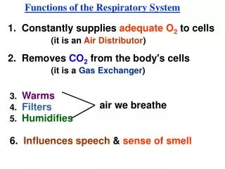
ONLY IN FEMALES’ SLIDES The Main Goal of Respiration Is to: 1- Provide oxygentotissues. 2- RemoveCO2 . يستحق تعطونه من وقتكم
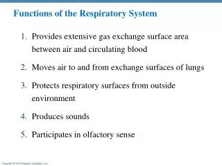
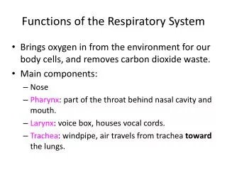
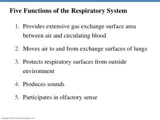
Other Functions of the Respiratory System • Gas exchange (respiratory function). • Phonation: the production of sounds by themovement of air through the vocal cords. • Pulmonary defense(protective functions): • Immunoglobulin A (Ig A). • Alpha-1 antitrypsin(protects the respiratory system from being ingested by trypsin). • Thepulmonary macrophagein the alveoli: engulfsmaller particles which pass through themuco- ciliary barrier filter. • Angiotensin* I is converted to angiotensin II with the help of angiotensin converting enzyme formed by the lungs. • Regulating the acid- base status of the body by washing out extra carbon dioxide from the blood(via hyperventilation). • Secretion of important substances like surfactant. *Angiotensin protein is very important in blood pressure regulation (it tends to raise blood pressure)it also promotes the secretion of aldosterone (a corticosteroid hormone which stimulates reabsorption of sodium by the kidneys and therefore regulates water and salt balance). Angiotensin breaks up into angiotensin I (inactive) then it is converted into angiotensin II (active) by ACE.
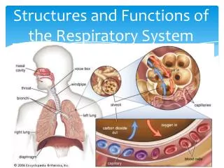
Respiratory Passages (Airways) * * * * * * * * * * *
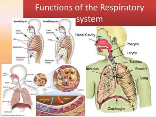
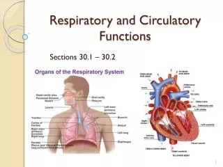
Respiratory Passages (Airways) Pulmonary capillaries for gas exchange The respiratory passage in general starts from the nose -> pharynx -> larynx -> trachea which divides into right and left bronchi and each one of them gives primary bronchi, secondary bronchi, tertiary bronchi and after these, there are terminal bronchioles and then respiratory bronchioles and then lastly the alveoli.
External & InternalRespiration • External respiration is gas exchange at the lung level. • Internal respiration is gas exchange at the tissue level. • Vessels which go to the heart (veins) • Vessels which travel from the heart (arteries) • Arteries always come out of ventricles. • The aorta travels from the left side of the heart carrying oxygenated blood to the body. External Respiration Respiratory unite in the lungs Pulmonary vein “carries oxygenated blood” Pulmonary artery “carries deoxygenated blood” Systemic artery “carries oxygenated blood” Systemic vein “carries deoxygenated blood” Internal Respiration
External & InternalRespiration (Extra notes from team 435) ** The pulmonary arteries are: deoxygenated blood, and the pulmonary veins are: Oxygenated blood. ** The systemic arteries are: Oxygenated blood, and the systemic veins are deoxygenated blood.
External & InternalRespiration • Respiration could beeither: • Resting: normal breathing during restingconditions. • Forced:(maximal): during exercise, in patientswithasthma,allergy, or any other pulmonary problems. *In some references (such as Guyton); regulation and ventilation is considered to be the 4th function of the respiratory system.
Lining Cells of The Alveoli Extra picture: for understanding the respiratory membrane. Respiratory membrane = the layers crossed by gas, type I pneumocytes, and basement membrane of the cells and the capillaries, and endothelial cells lining the capillary.
Surface Tension التوتر السطحي • H2O molecules at the surface are attracted to each other by attractive forces that resist distension called surface tension. • It is the attractive force between adjacent water droplets. • Surface tension increases (water droplets stick to each other) in contact with air. • Surface tension tends to oppose alveoli expansion. • Increased surface tension (water molecules are together) will constrict the alveoli. • In respiratory functions we decrease the surface tension of water molecules to be able to inflate the alveoli rather than collapsing them. • Pulmonary surfactant reduces surface tension. التوتر السطحي يسبب انكماش وتضيق الالفيولاي ودور السيرفكتنت انه يمنع تكون التوتر السطحي وبالتالي يساعد على سهولة زيادة حجم الالفيولاي ويصير التنفس وتبادل الغازات اسهل
Surfactant • Surfactant is a complex substance containing phospholipids and a number of apoproteins. • It lines the alveoli from the inside, which separates the air found in the lumen of the alveoli from water droplets on cells. • It is secreted by Type II alveolar cells. The earliest detection from fetal alveoli (of surfactant) begins between the (surfactant secretion starts at the) 6th-7th month (24th-28th week) but this could be delayed in others to week 35 of intrauterine life. • at week 35 surfactant is mature and begins to function. • Babies born before week 35 can be considered premature (before full maturation of surfactant) have immature surfactant, so the baby is vulnerable to alveolar collapse (RDS). • Full term pregnancy: 38 ∓2 weeks. • Premature babies: born between weeks 28 – 35 (approximately/no exact number). • Importance of surfactant is to reduces surface tension throughout the lung, prevents alveolar collapse, decreases airway resistance to inflationand decreases the work* of breathing and increases surface area for normal breathing. • Allergies increase airway resistance. • Work of breathing = the energy consumed during respiration. More about this topic in next slide. اي ممر للتنفس فالجسم لديه مقاومة بسيطة لمرور الهواء عبره، باعتبار ان الهواء جسم غريب والطبيعي تكون هناك مقاومة للاجسام الغريبة. المقاومة تتم عن طريق الـ Smooth musclesوهذا شيء طبيعي لكن، لو زادت انقباضات العضلات بالتالي يصير عندي increase airways resistance وهذا مايجعل مادة الـ Surfactant تقل. *How is surfactant related to the work of breathing? More surfactant -> less resistance -> air flows in easily -> with one breath a good amount of air flows in (e.g. 0.5 ml) -> less energy -> less work. Less surfactant -> more resistance -> air does not flow in easily -> you need more breaths to be able to reach the amount of air taken in by one normal breath (e.g. 0.5 ml) -> therefore putting in more energy -> more work.
Respiratory Distress Syndrome Innervations of Lungs and Bronchi Neonatal Respiratory Distress Syndrome: • Locally secreted factors: Histamine, slow reacting substances of anaphylaxis (SRSA) by mast cells, due to allergy (as in patients with asthma) often cause bronchiolar constriction and increased airway resistance. • They are substances produced locally by the mast cells in the lungs in response to irritation caused by entry of smoke or dust or any foreign antigens. • SRSA like bradykinin. (Anaphylaxis = allergy) Adult Respiratory Distress Syndrome: Smoking in adult, hypoxia or hypoxemia (low oxygen in the arterial blood) or both, decrease the secretion of surfactant and cause adult respiratory distress syndrome. All causes of adult respiratory distress syndrome decrease the secretion of surfactant, or destruct surfactant, which causes the syndrome. Also known as: Hyaline Membrane Disease. Deficiency in premature babies causes respiratory distress syndrome of the new born (RDS) which is a hyaline membrane disease. Here the surfactant is lacking. In the developing fetus. Infants born before week 24 will never have surfactant. Without surfactant, small alveoli have increased surface tension and increased pressures, and will collapse (atelectasis). Collapsed alveoli are not ventilated and therefore, cannot participate in gas exchange.
Link to Editing File (Please be sure to check this file frequently for any edits or updates on all of our lectures.) Quiz • https://www.onlinequizcreator.com/functions-and-organization-of-the-respiratory-system/quiz-249077 • References: • Girls’ and boys’ slides. • Guyton and Hall Textbook of Medical Physiology (Thirteenth Edition.)
Thank you! اعمل لترسم بسمة، اعمل لتمسح دمعة، اعمل و أنت تعلم أن الله لا يضيع أجر من أحسن عملا. The Physiology 436 Team: Female Members: ShroogAlsomali Alaa Alaqeel AseelAlsulaimani Laila Mathkour SondosAlhawamdeh ElhamAlami WateenAlhamoud Team Leaders: Qaiss Almuhaideb Lulwah Alshiha Male Members: Mohammed Alayed Contact us: Physiology436@gmail.com @Physiology436