Download
1 / 82
820 likes | 901 Views
Delve into the fascinating world of human vision and color perception, from the basics of light and color to the intricate workings of the human eye. Understand how light interacts with objects to create the colors we see and explore the complexities of color vision. Discover the structure of the human eye, the roles of rods and cones, and the pathways that process visual information in the brain. Gain insights into color terminology and perception, including hue, saturation, luminance, and more. Unravel the mysteries of color perception in this comprehensive exploration.

E N D
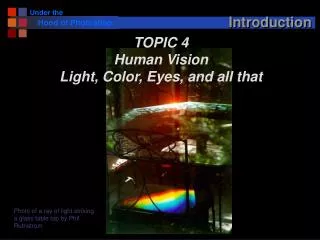
Introduction TOPIC 4 Human Vision Light, Color, Eyes, and all that Photo of a ray of light striking a glass table top by Phil Ruthstrom
Announcements • New course web page: • http://www-edlab.cs.umass.edu/cs391b/ • CS Saturday for juniors and seniors • http://www.cs.umass.edu/cs-saturday/ • NO CLASS: October 2 • Today: • More on the Photoshop histogram and fixing tonal problems • Start on human vision and color
Color Additive System Subtractive System
What’s Color? • It’s an attribute of an object (or thing) like texture, shape, smoothness • It depends upon • Spectral characteristics of the light illuminating the object • Spectral properties of the object (reflectance) • Spectral characteristics of the sensors of the imaging device (e.g. the human eye or a camera)
Light: EM Spectrum Electromagnetic Spectrum ‘Visible’ Spectrum
Newton 1666 From Voltaire's Eléments de la Philosophie de Newton, published in 1738
Spectral Distributions • Spectral distributions show the ‘amount’ of energy at each wavelength for a light source; e.g.
Interaction of Light and Matter • When light strikes an object, it will be • It will be wholly or partly transmitted. • It will be wholly or partly reflected. • It will be wholly or partly absorbed. • Physical surface properties dictate what happens • When we see an object as blue or red or purple, • what we're really seeing is a partial reflection of light from that object. • The color we see is what's left of the spectrum after part of it is absorbed by the object.
Spectral Reflectance Curves • Reflectance curves for objects that appear to be: The wavelengths reflected or transmitted from or through an object determine the stimulus to the retina that provokes the optical nerve into sending responses to our brains that indicate color.
The Human Eye Pupil - The opening through which light enters the eye - size from 2 to 8 mm in diameter Iris - The colored area around the pupil that controls the amount of light entering the eye. Lens - Focuses light rays on the retina. Retina - The lining of the back of the eye containing nerves that transfer the image to the brain. Rods - Nerve cells that are sensitive to light and dark. Cones - Nerve cells that are sensitive to a particular primary color.
Photoreceptor Low light receptors: ~125 million Color receptors: 5-7 million
Retinal Tissue LIGHT
Rods and Cones • Cones are located in the fovea and are sensitive to color. • Each one is connected to its own nerve end. • Cone vision is called photopic (or bright-light vision). • Rods give a general, overall picture of the field of view and are not involved in color vision. • Several rods are connected to a single nerve and are • Sensitive to low levels of illumination (scotopic or dim-light vision).
Absorption Curves Rods: achromatic vision The different kinds of cells have different spectral sensitivities Peak sensitivities are located at approximately 437nm, 533nm, and 610nm for the "average" observer.
Responses Response from i-th cone: si(l) = sensitivity of i-th cone t(l) = spectral distribution of light l = wavelength Cone sensitivity curves
Retina Cones in the fovea Moving outward from fovea Rods Cones Cones All of them are cones!
Retinal Processing 130 million sensors -> 10 million nerve fibers Processing at retinal level: center surround receptive fields This is what is sent down the optic nerve fibers
Illusions Center surround operators can be used to explain several ‘illusions’ Herring Grid Mach Bands
Visual Pathways • Past the eye, visual signals move through different processing stages in the brain. • There appear to be two main pathways • Magnocellular: low-resolution, motion sensitive, and primarily achromatic pathway • Parvocellular: high-resolution, static, and primarily chromatic pathway
Primary Visual Pathway Monocular Visual Field: 160 deg (w) X 175 deg (h)Binocular Visual Field: 200 deg (w) X 135 deg (h) Center Surround Orientation sensitive Motion sensitive Opponent Colors ..... FEATURES
Describing Color • Color is a very complex phenomenon • physical • psychological • Following description only skims the surface • important details omitted • simplified mathematics • ‘leaps of faith’
Terminology (Rough) • Hue: dominant wavelength of light entering the eye • Saturation: inversely proportional to amount of white light mixed with pure color • Red - fully saturated • pink - partially saturated • white - fully unsaturated • Luminance: intensity of light entering the eye • Lightness: luminance of a reflecting object • Brightness: luminance of a light source (radiance) • Chromaticity: hue and saturation (not luminance)
Brightness and Luminance • Question: What is the difference between luminance and brightness? • Answer:Luminance of an object is its absolute intensity. Brightness is its perceived luminance, which depends on the luminance of the surrounding. • Question:Why are luminance and brightness different? • Answer: because our perception is sensitive to luminance contrast rather than absolute luminance. Example: car headlights bother car driver much more at night (when it's dark) than in the day time. Luminance of headlights is the same, it's only the perceived luminance (brightness) that differs from night (dark) to daytime (light).
Brightness Adaptation • Range of light intensity levels to which HVS (human visual system) can adapt: on the order of 1010. • Brightness as perceived by the HVS is a logarithmic function of the light intensity incident on the eye. • The HVS cannot operate over such a range simultaneously. • For any given set of conditions, the current sensitivity level of HVS is called the brightness adaptation level.
Brightness Adaptation • The eye also discriminates between changes in brightness at any specific adaptation level. • Small values of Weber ratio mean good brightness discrimination (and vice versa). • At low levels of illumination brightness discrimination is poor (rods) and it improves significantly as background illumination increases (cones). • The typical observer can discern one to two dozen different intensity changes DIc: the increment of illumination discriminable 50% of the time and I : background illumination
Contrast vs. Intensity • We care about surface reflectance, not light intensity. • Contrast is proportional to reflectance. Intensity is reflectance*illumination Local contrast is c = (I-Imean)/Imean
Local Adaptation • Ellipses are the same gray level
Local Adaption • Ellipses are the same gray level
Observation of the Day The eye / brain combination is NOT a camera!
What Do We ‘See’? Light Sources Surface Reflectance Eye sensitivity
Reflectances….. Some Common Objects
Tristimulus Theory • Two light sources S1 and S2 may have very different spectral distribution functions and yet appear identical to the human eye. • The human retina has three types of receptors. • The receptors have different responses to light of different frequencies. • Two sources S1 and S2 will be indistinguishable if they generate the same response in each type of receptor. • same observer • same light conditions • called metamerism
Grassman’s Law (1835) • 1st Law: Any color stimulus can be matched exactly by a combination of three primary lights. • The match is independent of intensity • Basis of many color description systems • 2nd Law: adding another light to both of these stimuli changes both in the same way.
Color Matching Experiments Controllable standard sources - e.g. a, b, and g are user determined R a IR IG G b Controllable mix g B IB IR, IG.IB Ul Unknown color Monochromatic light of constant intensity Ul Following few slides adapted from Paul Avery, Univ. of Florida
Procedure • Upper part of field illuminated by adjustable monochromatic lights of wavelengths lR, lG, lB • lR = 645 nm, lG= 526 nm, lB = 444 nm • Lower part of field illuminated by a single monochromatic light of constant intensity Ul • Adjust RGB intensities until perfect match • Record intensities (IR, IG, IB) for that wavelength • Shift wavelengthl = l +Dl • Repeat What do we get?
Color Matching Functions • Recorded values of (IR, IG, IB) define color matching functions for the three light sources • If match requires negative value for one of the lights, add the light to the lower disk. Example: match unit intensity at 500 nm Use curves to get values IR=-0.30, IG=0.50, IB=0.10
Matching a spectrum • Any spectrum can be matched this way • break spectrum into n discrete samples • for each sample, calculate (Ri, Gi, Bi) as before • Add all (Ri, Gi, Bi) to get final (R, G, B) value • Simple!
CIE Color Matching Model • Problems: • Negative values • Difficult to deal with physically • Brightness not explicitly represented • 1920: Commission Internationale de l’Eclairage • (International Lighting Commission) • 1931: New Standard Color Model
CIE Color Model • Introduced three new (imaginary) primaries X, Y, Z so that all tristimulus values are positive • Can relate R, G, B to X, Y, Z mathematically, so no problem • Called x(l), y(l), z(l) functions XYZ values • Independent of initial choice of lR, lG, lB values!


