
WDR77 and Its Role in Histone Methylation: Insights into Epigenetic Regulation
This study explores the interactions of WDR77 with PRMT5 and modified histone H2A, shedding light on the specific substrates and functions of the WDR77/PRMT5 heterodimer complex. Post-translational modifications of histones regulate gene expression, and understanding the dynamics of WDR77 may clarify its role in cellular processes. The findings suggest potential mechanisms behind arginine methylation, focusing on the specificity of WDR77's recognition of histone modifications and its implications for genomic regulation and stem cell maintenance.

WDR77 and Its Role in Histone Methylation: Insights into Epigenetic Regulation
E N D
Presentation Transcript
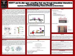
WDR77 and its alter egos: propelling their way through intracellular interactionsMadhumithaRengasamy1,2, SiDe Li 1,2, Martin J. Walsh1,2,3Departments of 1Structural and Chemical Biology, 2Pediatrics and 3Genetics and Genomic Sciences ABSTRACT Post-translational modification of histone tails is one of the primary modes of epigenetic regulation. The multiple types of post-translational modifications include acetylation, methylation, phosphorylation, ubiquitylation, sumoylation, ADP-ribosylation, prolineisomerization, citrullination, butyrylation, propionylation and glycosylation. Methylation of arginine residues in histone tails is a predominant type of modification and the marked feature of this modification is that different methylated states i.e. monomethylated or dimethylated can direct different transcriptional consequences. Protein argininemethyltransferases (PRMTs) are a group of enzymes that attach a methyl group to a guanidino nitrogen atom of arginine using S-adenosylmethionine (AdoMet) as the source of the methyl group. PRMT5 has been shown to have an intracellular dependence on the interacting WD40 repeat domain -containing protein WDR77 to determine specific substrates. Furthermore, the WD40 domain containing protein WDR77 has been shown to interact specifically with histone H2A. However, the dependence of the interaction between WDR77 and modified histone H2A has not been clarified. Some evidence suggests that H2AR3 methylation occurs in the cytosol and is deposited into nucleosomes by chaperones as a dimer with H2B, suggesting a sophisticated mechanism of nucleosome assembly and remodeling to ensure proper installation of H2AR3me2 throughout the genome. Furthermore, the role between PRMT5 and WDR77 to identify specific substrates remains unclear. In this study we begin to determine the criteria for WDR77 recognition of specific substrates for PRMT5 and to determine the functions of both cytosolic and nuclear isoforms of the WDR77/PRMT5 heterodimeric complex to enact specific genomic regulatory functions with H2AR3me2 methylation. Figure 1- Organization of the nucleosomalhistone NH terminus (Fullgrabe et. al. (2011) Oncogene 30:3391-3403). Figure 2- Distinction between assymmetrical and symmetrical argininedimethylation by protein argininemethyltransferases (PRMTs) (Di Lorenzo A. and Bedford M.T. (2011) FEBS Letters 585(13):2024-31) The WD40 repeat domain protein, WDR77 WD repeat containing protein 77 (WDR77) is a 342 aa WD repeat protein that is present in the nucleus and cytoplasm. It has seven putative WD domains, three Nuclear Localization Sequences (NLS) and two Nuclear Exclusion Sequences (NES). It has been separately reported as MEP50 (Methylosome protein 50) and p44 (co-factor of androgen receptor). Figure 4 - WDR77 interactions in the cytoplasm. WDR77/MEP50 is a part of the 20S methylosome complex. PRMT5/WDR77 complex catalyze the symmetric dimethylation of H2AR3 and this is essential for stem cell maintenance. Figure is not drawn to scale. Figure 5- WDR77 interactions in the nucleus. WDR77 interacts with proteins in the actively transcribing as well as repressed regions of the genome. AR/ER-androgen receptor or estrogen receptor, Cyc D1- Cyclin D1. Figure is not drawn to scale. Results Peptide pull down assay Nuclear Extract – PC3 nuclear extract Peptides used 1. No peptide – C 2. Unmethylated H3R2 peptide- UN 3. Assymmetricallydimethylated H3R2 peptide – 2A 4. Symmetrically dimethylated H3R2 peptide – 2S 2. Purification of WDR77 M-Marker FT- Flow through fraction W-Wash fraction E and E2- Elution fractions Cytoplasm Acid elution fractions Heat denatured fractions 20S Methylosome PRMT5 PRMT5 Me pICln WDR77 Sm proteins Nuclear membrane WDR77 H2A C UN 2A 2S C UN 2A 2S Nuclear Pore complex M WDR5 Sm proteins Nucleus H2AR3me2s WDR77 M H2AR3me2s/H2B dimer PRMT7 M FT W E E E2 FT W E E2 250 150 100 75 50 37 25 PRMT7 PRMT5 WDR77 P CycD1 WDR77 SUZ12 NuRD complex WDR77 WDR77 PRC-2 complex FCP1 PRMT5 Transcription MBD2 Pol II complex WDR77 WDR77 AR/ER Densely packed heterochromatin Conclusion Although the peptide pull down assay showed WDR77 interaction with the H3R2Me2s and H3R2Me2a peptides it remains to be confirmed if there is direct binding. The assay should be repeated using the purified WDR77 protein. Future experiments also include ChIP assays to determine WDR77 binding on a genome wide scale and crystallization studies to determine the structure of WDR77. mRNA Phosphate group Methyl CpG P WD40 proteins Among the many protein domains that recognize histone modifications, the group of WD repeat proteins constitute a large heterogenous family of proteins. The WD repeat proteins get their name by carrying repeats of amino acid residues within a stretch of 40-60 amino acids that begin with a Glycine-Histidine (GH) dipeptide and end in a Tryptophan-Aspartic acid dipeptide (WD). WD repeat generally contain 7 repeats that are arranged in a circular manner forming a β propeller structure (Figure 1). This structure enables WD repeat domains to act as a scaffold to accommodate the interaction and assembly with a diverse array of proteins and their structures. Figure 3 - H3K27me3 binding by EED (left and middle) and H3R2me2s binding by WDR5 (right), as shown from from crystal structures both at 1.9A. From Margureonet al. (2009) (left and middle) and Migliori et al. (submitted) (right). Aims To check if WDR77 recognizes and binds to specific histone modifications. Peptide pull down assay Biotinylated (bait) peptides Immobilized on streptavidin beads Incubated with nuclear extract Collect beads Remove bound proteins by acid elution/ denaturation by heat Western blot 2. Purification of WDR77 and check its binding with PRMT5 and PRMT7 Constructs (Ernesto lab IMCB, Singapore) WDR77-GST – pGEX6P1 PRMT7-GST – pGEX PRMT5-His- pET32a • References • Di Lorenzo, A. and M.T. Bedford, Histoneargininemethylation. FEBS Letters, 2011. 585(13): p. 2024-2031. • Friesen, W.J., et al., A novel WD repeat protein component of the methylosome binds Sm proteins. J BiolChem, 2002. 277(10): p. 8243-7. • Furuno, K., et al., Association of Polycomb group SUZ12 with WD-repeat protein MEP50 that binds to histone H2A selectively in vitro.BiochemBiophys Res Commun, 2006. 345(3): p. 1051-8. • Hosohata, K., et al., Purification and Identification of a Novel Complex Which Is Involved in Androgen Receptor-Dependent Transcription. Molecular and Cellular Biology, 2003. 23(19): p. 7019-7029. • Le Guezennec, X., et al., MBD2/NuRD and MBD3/NuRD, two distinct complexes with different biochemical and functional properties. Mol Cell Biol, 2006. 26(3): p. 843-51. • Licciardo, P., et al., The FCP1 phosphatase interacts with RNA polymerase II and with MEP50 a component of the methylosome complex involved in the assembly of snRNP. Nucleic Acids Res, 2003. 31(3): p. 999-1005. • Margueron, R., et al., Role of the polycomb protein EED in the propagation of repressive histone marks. Nature, 2009. 461(7265): p. 762-7. • Peng, Y., et al., Androgen receptor coactivator p44/Mep50 in breast cancer growth and invasion. Journal of • Cellular and Molecular Medicine, 2010. 14(12): p. 2780-2789. • 9. Tee, W.W., et al., Prmt5 is essential for early mouse development and acts in the cytoplasm to maintain ES cell pluripotency. Genes & Development, 2010. 24(24): p. 2772-2777. Acknowledgements I would like to thank Dr. Martin Walsh for his invaluable guidance. I would also like to thank all members of the Walsh lab for helpful discussions about experiments and their unstinted support and encouragement.