Download
1 / 31
310 likes | 347 Views
Explore a detailed case study of a 51-year-old male presenting with altered sensorium, neurological symptoms, and a complex medical history, including TB, DM, and CKD. The multidisciplinary medical team led by Prof. Dr. R. Balajinathan unravels the diagnostic puzzle and devises a treatment plan including IV fluids, antibiotics, and neurosurgical interventions.

E N D
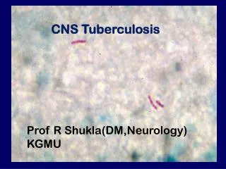
INTERESTING CASE OF CNS TUBERCULOSIS II MEDICINE UNIT PROF DR.R .BALAJINATHAN MD., ASST PROF DR.V.N.ALAGAVENKATESAN MD., DR.P.V BALAMURUGAN MD. DR.R.PANDISELVAM M.D
CASE 2 51 years old male admitted with chief complaints of • Altered sensorium for past 3 days. HISTORY OF PRESENTING ILLNESS He was apparently normal 3 months back after which he developed low back pain , fever with chills and rigor, was diagnosed to have pyelonephritis and treated with DJ stenting. • H/O difficulty in using right upper and lower limb past 1 wk • H/O difficulty in walking • H/O Headache • H/O altered sensorium – 3 days • H/O fever Intermittent in nature Associated with chills & rigor • H/O loss of appetite • H/O loss of weight
No H/O seizures • No H/O vomiting • No H/O LOC • No H/O head injury • No H/O involuntary micturition/ defecation • No H/O cough with expectoration • No H/O breathing difficulty • No H/O ear pain/ discharge in the ear • No H/O blurring of vision
PAST HISTORY: • K/C PTB 6 years back completed 6 months of ATT • K/C of pyelonephritis underwent DJ stenting 3 months back • K/C of TYPE 2DM on OHA • Not a k/c of SHT/ CAD/Seizure disorder PERSONAL HISTORY: • Consumes mixed diet • Known smoker & alcoholic 15 years
GENERAL EXAMINATION • Patient drowsy occasionally responds to painful stimuli • Afebrile • Anaemic • Not icteric • No cyanosis • No clubbing • No lymphadenopathy • No pedal oedema
VITAL SIGNS: PULSE : 86/ min regular in rhythm normal in volume felt in all accessible peripheral pulse • No radiofemoral ,radio radial delay • BLOOD PRESSURE: 150/90 mmhg taken in right upper limb in supine position • SPO2 : 94% @ room air • RR : 14 / min
SYSTEMIC EXAMINATION CENTRAL NERVOUS SYSTEM HMF - Drowsy , occasionally responds to oral commands Language – Could not be tested Speech Memory
CRANIAL NERVE EXAMINATION • I – could not be tested • II - B/L PUPIL 3MM ERTL FUNDUS – NORMAL • III , IV, VI- No apparent ptosis • V- No jaw deviation Jaw jerk just present • VII -No facial asymmetry No deviation of angle of mouth Wrinkles + Eye lid closure full • VIII, IX,X,XI, XII- could not be tested
CNS EXAMINATION • SENSORY SYSTEM • POSTERIOR COLUMN Could not be tested • CEREBELLUM • SPINE & CRANIUM - Normal
SYSTEMIC EXAMINATION • CVS – S1S2 + JVP Normal NO Murmur • RS B/L AE + NVBS No added sounds • PA Soft No organomegaly, BS +
INVESTIGATION • Hb 10.8 gm • TC 16900 cells/ cumm • DC P- 82 L – 11 M- 7 • PLT 3.25 lakhs • PCV 34% • ESR 31 mm/hr • RBS 422 mg/dl • LFT Normal
INVESTIGATIONS… URINE ANALYSIS • ALBUMIN - TRACE • SUGAR - NIL • DEPOSITS- 2-4 PUS CELL • URINE KETONES - Negative • USG ABDOMEN- Medical renal disease • VCTC - NON REACTIVE • BLOOD & URINE C&S - No evidence of growth in culture
CT BRAIN REPORT • Mixed lesion noted in left temperoparietooccipital region causing midline shift with obstruction of third ventricles. • IMPRESSION ; SPACE OCCUPYING LESION in the left temperoparieto occipital region • SUGGESTION MRI BRAIN
DIAGNOSIS • TYPE 2 DM • SOL LEFT TEMPERO PARIETAL REGION WT RIGHT HEMIPLEGIA • OLD PULMONARY TUBERCULOSIS • CHRONIC KIDNEY DISEASE
TREATMENT • IV fluids • Inj. Dexamethasone 8mg iv bd • Inj ceftriaxone 2g iv bd • Inj ampicillin 2g iv qid • Inj ranitidine 50 mg iv bd • Glycerol 20ml tds by RT • Inj Phenytoin 100mg iv tds • Inj Human actrapid 8units tds
NEUROLOGIST OPINION • Right hemiplegia / SOL – Left Temparo Parietal Region With Midline Shift SUGGESTION • Oral Glycerol 20ml tds • Inj Phenytoin 100 mg IV TDS • Inj dexamethasone 4mg IV BD • Neurosurgeon opinion • MRI Brain
NEUROSURGEON OPINION Right hemiplegia with aphasia S/O Glioblastoma Multiforme SUGGESTION • IVF NS 3PINT RL 2 PINT @ 100ml/hr • Head End Elevation • Inj Ceftriaxone 2g Iv Bd • Oral glycerol 20 ml TDS • Inj Phenytoin 100 mg IV TDS • Inj Lasix 20 Mg Iv Bd • MRI Brain with DWI Plain & Contast
MRI BRAIN REPORT • Multiple well defined T2 FLAIR hyperintense with hypointense lesion • T1 hyperintense with hypointense rim noted in left temperoparieto occipital region which shows diffusion restriction on DWI with extensive perilesionalvasogenic white matter edema noted • Lesion causing mSass effect in the form of midline shift of 15mm • Mass effect causing effacement of I/L lateral ventricle & temporal horn with dilatation of C/L lateral ventricle & temporal IMPRESSION; Multiple cerebral abscess - Tuberculous etiology Suggested contrast study
DIAGNOSIS • Type 2DM • CKD • OLD PTB • MULTIPLE CEREBRAL ABSCESS TB ETIOLOGY
NEUROSURGEON REVIEW • MULTIPLE CEREBRAL ABSCESS SUGGESTION; • To continue antiepileptics • To continue antiedema measures • To continue higher antibiotics
PULMONOLOGIST H/O PTB 6 years back , took ATT for 6 months SUGGESTION • Start on CAT II ATT • Ethambutol 600mg on alternative days • Inj. Streptomycin 500 mg on alternative days
AIM OF PRESENTATION • Rarity of CNS TB presenting as abscess • This case highlights the diagnostic dilemma in an immunocompromised patient with multiple focal brain lesions, especially in areas where TB is endemic. • TB should be considered as a possible aetiology for any intracranial lesion with radiological appearance suggestive of an abscess and surgical treatment can be kept as an option in case of neurological deficits, hydrocephalus or treatment failure.