Download
1 / 62
880 likes | 1.3k Views
Protein Structure. Protein Structure I. Primary Structure. Primary Structure Insulin. Bovine: Insulin. Figure 5-1. Human: ProInsulin. Signal sequence Chain B MALWMRLLPLLALLALWGPDPAAA FVNQHLCGSHLVEALYLV C Peptide CGERGFFYTPKT RREAEDLQVGQVELGGGPGAGSLQPLALEG Chain A

E N D
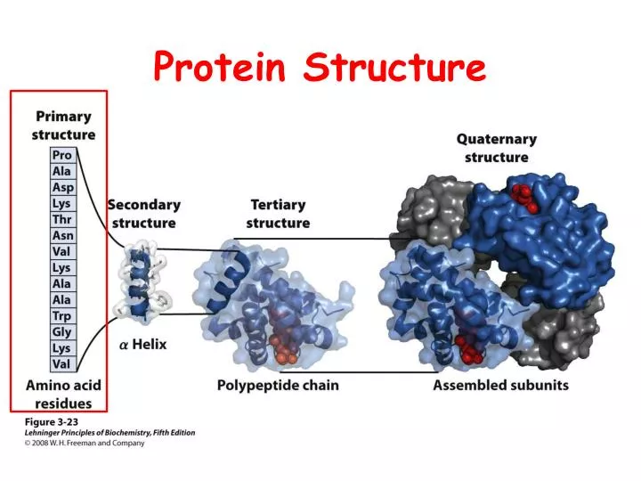
Protein Structure I Primary Structure
Primary Structure Insulin Bovine: Insulin Figure 5-1 Human: ProInsulin Signal sequence Chain B MALWMRLLPLLALLALWGPDPAAAFVNQHLCGSHLVEALYLV C Peptide CGERGFFYTPKTRREAEDLQVGQVELGGGPGAGSLQPLALEG Chain A SLQKRGIVEQCCTSICSLYQLENYCN
Primary Structure Insulin Bovine: Insulin Figure 5-1 Human: ProInsulin Signal sequence Chain B MALWMRLLPLLALLALWGPDPAAAFVNQHLCGSHLVEALYLV C Peptide CGERGFFYTPKTRREAEDLQVGQVELGGGPGAGSLQPLALEG Chain A SLQKRGIVEQCCTSICSLYQLENYCN
Value of Primary Structure Information • Primary sequence information is • prerequisite for determining three-dimensional structure • essential in understanding molecular mechanism of action • Sequence comparisons among analogous proteins • provide insights into protein function • reveal evolutionary relationships • Sequence of proteins whose mutations result in inherited diseases • assist in development of diagnostic tests • assist in development of effective therapies
Strategy • Purification of protein to homogeneity • Prepare protein for sequencing • Sequence polypeptide chains • Organize completed structure Alternative: Nucleic Acid Sequencing
Sequencing StrategySummary Figure 5-12
Sequencing Strategy I Figure 5-12
Sequencing Strategy II Figure 5-12
Sequencing Strategy III Figure 5-12
Prepare Protein for Sequencing • End Group Analysis: How many different subunits • Cleavage of disulfide bonds • Separation and purification of the polypeptide chains • Amino acid composition
End Group Analysis(How Many Different Subunits?) N-Terminal Identification
End Group Analysis(How Many Different Subunits?) C-Terminal Identification
Sequence Polypeptide Chains • Specific peptide cleavage reactions • Separation and purification of peptide fragments • Sequence determination
Hydrolysis Hydrolysis Polypeptide Amino Acids
Problems • Complete destruction of Trp • Partial destruction of Ser, Thr, and Tyr • Deamination of Asn and Gln
Enzymatic Hydrolysis Mild Conditions Many proteases and peptidases Specific and non-specific Problem: contribution of amino acids from hydrolysis of proteases
Amino Acid Analysis(Automated) Ion-exchange chromatography High performance liquid chromatography Colorimetric Analysis
Proteolytic Enzymes Cleave peptide bonds Specificity: R1
Specificity of Endopeptidases Table 5-3
Electrospray Ionization Mass Spectrometry (ESI) Figure 5-16a part 1
Electrospray Ionization Mass Spectrometry (ESI) Figure 5-16a part 2
Electrospray Ionization Mass Spectrometry (ESI) Figure 5-16b
Tandem Mass Spectrometry Figure 5-17
Organize Completed Structure • Ordering peptide fragments • Assignment of disulfide bond positions • Determine position of amides