Download
1 / 22
280 likes | 850 Views
PATIENT POSITIONING IN NEUROANAESTHESIA. Dr. Rahul Norawat. University College of Medical Sciences & GTB Hospital, Delhi. Pathophysiology Neg. venous pressure & exposure of veins & boney venous sinuses.

E N D
PATIENT POSITIONING IN NEUROANAESTHESIA. Dr. RahulNorawat University College of Medical Sciences & GTB Hospital, Delhi
Pathophysiology • Neg. venous pressure & exposure of veins & boney venous sinuses. • Surgical site is exposed to air & located above the level of heart, air may entrained in the veins & sinuses. • Consequences depends on volume, rate of entry, pt position, use of N2O & PFO (10-25% incidence) • Large VAE may CO by creating airlock & Lt. ventricular output.
Critical volume of air: 200-300 ml or 3-5 ml/kg. “The closer the vein of entrainment to the Rt. heart the lower the critical volume.”
Detection Laboratory Studies • Neither sensitive nor specific. • Routine lab tests to evaluate the associated end organ injury. • ABG : • Hypoxemia, • Hypercapnia, • Metabolic acidosis secondary to R-L pul shunting.
Imaging Studies • TEE most sensitivity. detect 0.02 ml/kg of air. • Added advantage of identifying PAE. • Precordial Doppler USG most sensitive noninvasive method. detect as little as 0.05 ml/kg of embolized air. • Incidence of VAE in sitting position, • 20-50% with precordial Doppler, • 76% with TEE, more sensitive.
Transcranial Doppler USG commonly used to detect cerebral microemboli. • CT detect VAE in axillary, subclavian veins, Rt. ventricle & pul. art. detect >1 ml air, specificity is best with large filling defects. • MRI show water conc. in affected tissues, not reliable. • CXR normal or may show gas in the pul art system, pul art dilatation, focal oligemia (Westermark sign) & pul edema.
Other Tests • ECG - Low sensitivity. resemble venous thromboembolism • Tachycardia, • Rt ventricular strain pattern, • ST depression. • EtCO2 - Change in 2 mmHg EtCO2 is indicator. nonspecific can occur with PE, massive blood loss, hypotension, circulatory arrest, upper airway obstruction.
EtN2 - Most sensitive gas-sensing VAE detection modality. • Pulse oximetry - Late findings, SpO2. • Pul. artery catheter - Detect, PAP secondary to mechanical obstruction/vasoconstriction from the hypoxemia induced by VAE. insensitive monitor (0.25 ml/kg). • Central venous catheter - Aspiration of air. CVP. • Esophageal Stethoscope - low sensitivity, detect “mill wheel murmur” (1.7 ml/kg/min).
Management • Notify surgeon asap. • Administer 100% O2, • Flood the field with saline, apply bone wax, • Turn off nitrous oxide, • Bilateral IJV compression, • Avoid hypotension - vasopressors (ephedrine).
Place patient in left lat decubitus & Trendelenburg position (Durant position). • Aspiration of entrained air- multi or single orifice catheter placed at the high level of right atrium. • PEEP: cerebral VP, • G-suit, • Supportive therapy & fluid resuscitation.
Initiate CPR - Maintaining CO, break large air bubbles into smaller and force air out of the rt ventricle into the pul vessels. • Hyperbaric oxygen therapy (HBOT) • Indications: Neurological manifestations and cardiovascular instability. • Benefits: • Compression of existing bubbles, • Establishing a high diffusion gradient to speed resolution of existing bubbles, • Improved oxygenation of ischemic tissues, • Lowered ICP.
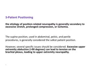
Summary of Task Force Consensus on the Prevention of Perioperative Peripheral Neuropathies Relevant to Positioning for Neurosurgery . Preoperative assessment • Ascertain that patients can comfortably tolerate the anticipated operative position.
Upper extremity positioning • Arm abduction should be limited to 90° in supine patients; patients who are positioned prone may comfortably tolerate arm abduction >90°. • Position arms to decrease pressure on ulnar groove (humerus). When arms are tucked at the side, neutral forearm position is recommended. When arms are abducted on armboards, either supination or a neutral forearm position is acceptable. • Prolonged pressure on the radial nerve in the spiral groove of the humerus should be avoided. • Extension of the elbow beyond a comfortable range may stretch the median nerve.
Lower extremity positioning • Prolonged pressure on the peroneal nerve at the fibular head should be avoided. • Neither extension nor flexion of the hip increases the risk of femoral neuropathy. Protective padding • Padded armboards may decrease the risk of upper extremity neuropathy. • The use of chest rolls in laterally positioned patients may decrease the risk of upper extremity neuropathies. • Padding at the elbow and at the fibular head may decrease the risk of upper and lower extremity neuropathies, respectively.
Equipment • Properly functioning automated BP cuffs on the upper arms do not affect the risk of upper extremity neuropathies. • Shoulder braces in steep head-down positions may increase the risk of brachial plexus neuropathies. Postoperative assessment • A simple postoperative assessment of extremity nerve function may lead to early recognition of peripheral neuropathies.
Documentation • Charting specific position actions during the care of patients may result in improvements of care by helping practitioners focus attention on relevant aspect of patient positioning; and providing information that continuous improvement processes can use to lead to refinement in patient care.
References • Miller RD. Anesthesia. 7th ed. NY: Churchill Livingstone Inc.; 2010. • Clinical Anaesthesia, Barash, Cullen & Stoelting, 5thed. • Rozet I, Vavilala S. Risks and Benefits of Patient Positioning During Neurosurgical Care : Anesthesiol Clin. 2007 Sept; 25(3): 631-62. • American Society of Anesthesiologists. Task Force on the Prevention of Perioperative Peripheral Neuropathies: Practice Advisory for the Prevention of Perioperative Peripheral Neuropathies. Anesthesiology 2000;92: 1168–1182. • American Society of Anesthesiologists. Mirski A etal, Diagnosis and Treatment of Vascular Air Embolism: Anesthesiology 2007; 106:164–77.