Download

1 / 16
160 likes | 274 Views
Spontaneous Oscillations From In Vitro Slices of Rat Neocortex. Clara Boyd, Lucia Chemes, Alberto Lopez, Angelos Stavrou. Reports of Spontaneous Activity In the Brain. EEG recordings in humans (awake & asleep) show oscillations in the absence of sensory or motor

E N D
Spontaneous Oscillations From In Vitro Slices of Rat Neocortex Clara Boyd, Lucia Chemes, Alberto Lopez, Angelos Stavrou
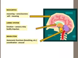
Reports of Spontaneous Activity In the Brain • EEG recordings in humans (awake & asleep) show oscillations in the absence of sensory or motor stimulation (10 Hz – 80 Hz) • Slices of mammalian neocortex maintained in vitro display slow oscillations (< 5 Hz)
Functional Relevance • Learning & Memory - storage & retrieval of neuronal activity patterns • Sensory-Motor Integration - replay of activity patterns during sleep • Neural Circuit Development - spontaneous activity in embryonic retina & thalamus before any visual experience • Regeneration & Repair Following Injury - synchronous neuronal activity is a signal for axonal sprouting after cortical lesions in the adult
Cellular Mechanisms • Developmental Ca+2 waves mediated by gap junctions in developing neocortex (IP3 second messenger) • Intrinsic (Pacemaker Connections) persistent Na+ current & hyperpolarization- activated cation current • Synaptic-Contact Mediated (Network Connections) activation of GABAergic & glutamatergic synapses
Observing Activity In Vitro: Method • Slice Preparation (area V of neocortex, 300m thick) • Loading with Ca+2 indicator Fura-2AM & imaging via 2-Photon Microscopy Correlation between Action Potentials and Somatic Ca+2 Transients Relationship between action potentials of single neuron & population state of the network
Image Analysis: defining neurons • Image enhancement for visual analysis • Definition of neuron • Looking for circle-shaped objects around 10m diameter • Imaging performs a slice, so neurons can be cut and sizes may vary • Problems • Neurons have deferent shapes and sizes • Background is not uniform and contains calcium traces from dendrites • Neurons change ‘shapes’ in time
Image Analysis: extracting neurons threshold Enhanced image cleaning separation
Image Analysis: tracking neurons in time • Neurons recognition have problems • Some neurons may split, merge, appear and disappear because of the recognition algorithm (noise) • They may be recognized as new neurons every frame Shape change split merge • The tracking algorithm takes car of this issues, recognizing all those problems and tracking neurons even if the recognition is not perfect • Tracks exact shape every frame, reducing noise
Statistical Correlation of the Spike Trains Numberof pairs Correlation of pairs
Future Directions • Obtain Data with higher sampling ratio (at least 4-5Hz. • Create a spacial mapping of the correlated neurons in the image. • Being able to predict according to the spatial position of the neuron the probability of being connected to another neuron for different areas of the brain. • Use the same image analysis in different layers of a 3-D image and predict with greater accuracy the connection. • Repeat the experiment with different images and test the filters and the thresholds already implemented.