Download
1 / 5
50 likes | 53 Views
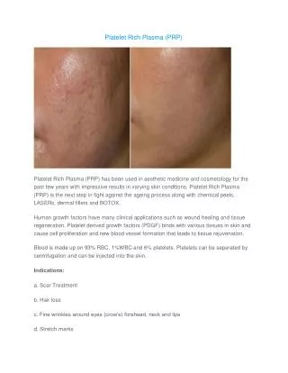
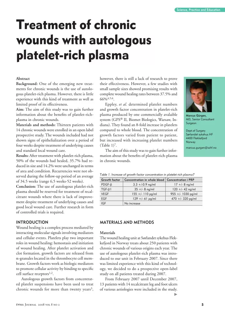
PRP treatment is used to treat a wide variety of chronic diseases. Here's the study.<br><br>Source: https://medgenerx.com/treatment-of-chronic-wounds-with-autologous-platelet-rich-plasma/

E N D
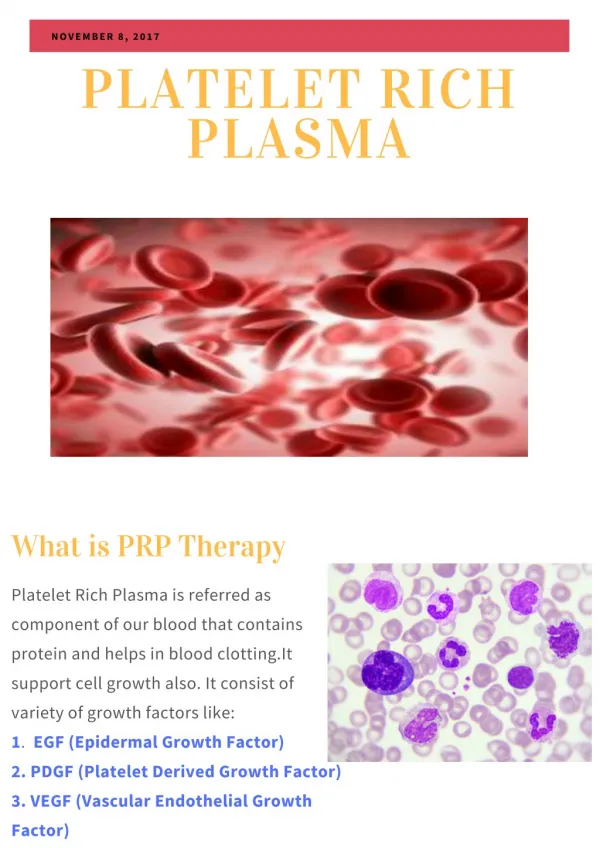
Science, Practice and Education Treatment of chronic wounds with autologous platelet-rich plasma Abstract Background: One of the emerging new treat- ments for chronic wounds is the use of autolo- gous platelet-rich plasma. However, there is little experience with this kind of treatment as well as limited proof of its effectiveness. Aim: The aim of this study was to gain further information about the benefits of platelet-rich- plasma in chronic wounds. Materials and methods: Thirteen patients with 14 chronic wounds were enrolled in an open label prospective study. The wounds included had not shown signs of epithelialization over a period of four weeks despite treatment of underlying causes and standard local wound care. Results: After treatment with platelet-rich plasma, 50% of the wounds had healed, 35.7% had re- duced in size and 14.2% were unchanged in terms of area and condition. Recurrencies were not ob- served during the follow-up period of an average of 34.5 weeks (range 6,5 weeks-52 weeks). Conclusion: The use of autologous platelet-rich plasma should be reserved for treatment of recal- citrant wounds where there is lack of improve- ment despite treatment of underlying causes and good local wound care. Further research in form of controlled trials is required. however, there is still a lack of research to prove their effectiveness. However, a few studies with small sample sizes showed promising results with complete wound healing rates between 37.5% and 66%4,5,6. Eppley, et al. determined platelet numbers and growth factor concentration in platelet-rich plasma produced by one commercially avalaible system (GPS® II, Biomet Biologics, Warsaw, In- diana). They found an 8-fold increase in platelets compared to whole blood. The concentration of growth factors varied from patient to patient, but increased with increasing platelet numbers (Table 1)7. The aim of this study was to gain further infor- mation about the benefits of platelet-rich-plasma in chronic wounds. Marcus Gürgen, MD, Senior Consultant Surgeon Dept.of Surgery Sørlandet sykehus HF 4400 Flekkefjord Norway marcus.gurgen@sshf.no Table 1. Increase of growth factor concentration in platelet rich plasma7 Growth factor Concentration in whole blood Concentration i PRP PDGF-b 3.3 +/-0.9 ng/ml TGF-b1 35 +/- 8 ng/ml VEGF 155 +/- 110 pg/ml EGF 129 +/- 61 pg/ml IGF No increase 17 +/- 8 ng/ml 120 +/- 42 ng/ml 955 +/- 1030 pg/ml 470 +/- 320 pg/ml INTRODUCTION Wound healing is a complex process mediated by interacting molecular signals involving mediators and cellular events. Platelets play two important roles in wound healing: hemostasis and initiation of wound healing. After platelet activation and clot formation, growth factors are released from a-granules located in the thrombocyte cell mem- brane. Growth factors work as biologic mediators to promote cellular activity by binding to specific cell surface receptors1,2. Autologous growth factors from concentrat- ed platelet suspensions have been used to treat chronic wounds for more than twenty years3, MATERIALS AND METHODS Materials The wound healing unit at Sørlandet sykehus Flek- kefjord in Norway treats about 250 patients with chronic wounds of various origins each year. The use of autologous platelet-rich plasma was intro- duced to our unit in February 2007. Since there was limited experience with this kind of technol- ogy, we decided to do a prospective open-label study on all patients treated during 2007. From February 2007 until December 2007, 13 patients with 14 recalcitrant leg and foot ulcers of various aetiologies were included in the study. EWMA Journal 2008 vol 8 no 2
Figure 2: Platelet concentrate and thrombin form a clot when mixed in a sterile container. Figure 3: The clot can be cut according to wound size before being transferred to the wound. Figure 1: The GPS-II-tube after spinning. Preparation of platelet-rich plasma Platelet-rich plasma gel to treat wounds was prepared by using a desktop centrifugation system (Gravitational platelet separation system, GPS® II, Biomet Biologics Inc., Warsaw, Indiana) and a reaction chamber to pro- duce autologous thrombin (Thrombin Processing Device, TPD™, Thermogenesis Corp., Rancho Cordova, Cali- fornia). Making platelet-rich plasma starts by drawing a volume of 55 ml blood from the patient and mixing it with 5 ml citrate. The blood is then transferred to the GPS®- disposable which is placed in a centrifuge. It is spun at 3200 rpm for 15 minutes. During centrifugation, blood is separated into three different fractions: platelet-poor plasma, platelets and white blood cells, and red blood cells (Figure 1). The platelet-poor plasma and the concentrated platelets and white blood cells are drawn from the tube using separate ports. Thrombin is produced by mixing platelet-poor plasma with an ethanol/CaCl2-reagent in the TPD™ reaction chamber. Platelet concentrate and thrombin are then drawn into seperate syringes, the plate- let-thrombin ratio at 10:1. The syringes are connected using a Y-connector. When mixed, thrombin activates the platelets. The result is a platelet gel that sticks to the sur- face of the wound when applied. Growth factors are also released upon platelet activation. Three different methods can be used to apply platelet gel onto wounds. Using the Y-connector, thrombin and platelet concentrate can be sprayed as a steady stream into the wounds. This method fits best to fill cavity wounds. It is also possible to attach a special tip to the Y-connector, which applies the mixture as a fine spray onto more shallow or large wounds. A third possibility is to create a clot in a sterile container, and then transfer the clot as a whole or cut into pieces into the wound (Figure 2, 3). As more experience with this type of treatment was aquired, the latter was preferred due to less leakage, more stable clotting and better utilization of the amount of platelet gel. The sample consisted of 3 females and 10 males with an average age of 52.1 years (range 35-76). The largest groups of wound diagnoses were venous leg ulcers (n=6) and dia- betic foot ulcers (n=3). The average duration of the ulcers was 6.8 years (range 2 months-21 years). Since treatment with platelets is regarded as an estab- lished method, acceptance from the local ethical commit- tee was not needed. All patients had given their written informed consent before entry to the study. Methods This was a prospective open label study to look at the effects of treatment with autologous platelet-rich plasma. Inclusion criteria were chronic wounds with a dura- tion more than 8 weeks that had not shown any progress (decrease in size, formation of granulation tissue, epitheli- alization) over a four-week period despite treatment of the underlying causes and appropriate local wound treatment. Wounds had to be free for necrosis. The ankle-brachial pressure index (ABPI) had to be more than 0.6. Ulcers that showed evident clinical signs of infection or were exudating heavily, were excluded. Determination of ankle-brachial pressure index was done on all patients. Venous leg ulcers were assessed by clinical features and by measuring the ankle-brachial pressure index . All diabetic ulcers were assessed with both ankle-bra- chial pressure and toe pressure. An ABPI < 0,8 and toe pressures less than 30-50 mmHg indicated arterial disease. Neuropathy was examined by testing sensibility in diabetic feet with a 10-g-Semmes-Weinstein-monofilament and a 128 MHz-tuning fork. Wounds were also assessed in terms of formation of granulation tissue, moisture balance, and infection. These assessments were used to describe the wounds as improved, unchanged or deteriorated. EWMA Journal 2008 vol 8 no 2
Science, Practice and Education Figure 4: Patient nr. 2 (Table 2). A neuroischemic diabetic heel ulcer prior to treatment with platelet gel. Figure 5: Day 27: the wound size is reduced by 40%. RESULTS Wounds were measured at day 0 with digital planimetry and showed an average wound size of 6.7 cm2 (range 0.4 cm2-22.3 cm2). On day 7 after treatment, ulcer size had reduced by an average of 31.4% (range 2.1%-77.7%) in 11 of 14 wounds. Two wounds were unchanged in size, and 1 wound had increased in size by 4.3%. 13 wounds were clinically assessed as improved, and 1 as unchanged. After 28 days, 1 wound had healed completely. Of the remaining ulcers, 12 had decreased in size to an average of 55.2% (range 6.2%-80%) of their original size. All of those were clinically assessed as improved. One wound remained unchanged in both size and condition. The number of treatments with platelet-derived growth-factors varied from 1-4 (average 2.1 treatments). Follow-up continued for an average of 8.4 months (range 1.5-12). Two ulcers showed signs of infection dur- ing the course of treatment. In both cases, Pseudomonas species were cultured and infections successfully treated with systemic antibiotics. Other side-effects were not re- corded. Seven (50%) of the ulcers healed within an average of 153 days (range 30-317). Of the non-healed wounds, 2 (13.3%) showed signs of improvement within the first 4 weeks, but have since deteriorated. The remaining 5 (35.7%) of the wounds continued to show improvement and are between 68.7% and 6.8% of their original size. (Table 2) Recurrencies were not observed. Figure 6: Day 155: The wound has healed after two applications of platelet gel, off-loading and good local wound care. A degradable dressing (Topkin, Biomet Europe, Dor- drecht, the Netherlands) and a secondary absorbent layer (Mepilex, Mølnlycke Health Care, Gothenborg, Sweden) were used to cover the wounds. Treatment of the underly- ing wound causes such as compression therapy for venous ulcers and off-loading for diabetic ulcers was continued. Outcome measures In order to assess wound healing accurately, we used dig- ital planimetry (Visitrak, Smith & Nephew, Hull, UK) to measure wound sizes. Digital measurements of the ulcers size were routinely taken on day 7 and day 28. As wounds were followed-up, wound tracings were performed about every four weeks. The primary endpoint was time to healing, and the secondary endpoint was reduction in ulcer size if wounds had not healed. DISCUSSION Wound healing is a complex process that is regulated by interactions between a large number of cell types, extracel- lular matrix proteins and mediators such as cytokines and growth factors. Lack of balance between these interactions may result in a chronic wound. One possible cause of imbalance in the wound healing process is high bacterial EWMA Journal 2008 vol 8 no 2
Science, Practice and Education Table 2. Patient characteristics Nr. Sex, age Wound diagnosis Duration of the wound 10 years Wound size (cm²) Number of treatments %-change in area at day 7 22.5 %-change in area at day28 27.1 Result December 2007 1 F, 76 Venous leg ulcer 21.8 1 Deteriorated, no signs of epithelialization Healed day 155 Healed day 30 Healed day 62 2 3 4 M, 53 F, 64 M, 56 Neuroischemic diabetic heel ulcer Neuroischemic diabetic heel ulcer Traumatic leg ulcer associated with an open distal leg fracture Pressure ulcer, leg, associated with spinal cord injury Venous leg ulcer Venous leg ulcer Venous leg ulcer Mixed-etiology leg ulcer 27 weeks 30 weeks 8 weeks 22.3 0.9 4.8 2 1 1 23.3 77.7 2.1 49.7 100 16.6 5 M, 39 1 year 5.6 4 7.7 16.1 Reduced by 42.9% 6 7 8 9 M, 50 F, 72 M, 44 M, 55 18 years 21 years 30 weeks 4 years 2.1 1.3 4.4 16.3 4 2 3 1 0.0 31.8 + 4.3 14.2 0.0 69.3 13.1 38.7 Healed day 233 Healed day 238 Reduced by 93.2% Deteriorated after 10 weeks Reduced by 59.7% Reduced by more than 80% (impossible to meas- ure due to small size) Healed by day 317 Reduced by 72% / healed by day 35 10 11 M, 39 M, 35 Venous leg ulcer Heel ulcer after cutaneous flap transfer 3 years 21 years 5.7 0.7 4 2 0.0 57.1 6.2 71.4 12 13 M, 58 M, 37 Neuropathic diabetic heel ulcer Venous leg ulcers 36 weeks 9 years 0.4 1 1 50 50 8.0 / 1.5 12.5 / 46.6 68.8 / 80 in in both venous and diabetic ulcers13,14,15,16. Crovetti, et al., McAleer, et al., and Steenvorde, et al. reported wound closure rates in recalcitrant ulcers between 37.5% and 66%4,5,6. The results of this small sample of patients show that 50% of all wounds healed. Treatment with platelet-rich plasma was reserved for patients with wounds that had not healed despite the use of other treatment strategies. Some of these prior treatments lasted for many years as the average duration of ulcers treated in this study (6.8 years) shows. In those cases, the use of platelet-rich plasma not only lead to wound closures, but also to improved the healing time. The average healing time for the wounds that closed was less than 6 months. 42,8% of 7 venous ulcers in this study healed within 6 months after treatment with platelet-rich plasma and two- layer compression bandage. Nelson, et al. showed 67% healing rates of venous ulcers within 24 weeks when us- ing four-layer compression therapy and 49% healing rates when using single-layer compression therapy17. However, it might be difficult to compare outcomes because of the low number of patients included in the present study. Two minor complications in form of infection with Pseudomonas aeruginosa were observed. Both wounds were cultured for Pseudomonas aeruginosa prior to plate- let gel treatment. In both cases, platelet-rich plasma was sprayed on the wounds. The clot that forms in the wound after spraying does not seem to be as stable as the clot produced in a sterile container. There is also some leakage when spraying, which may contribute to an excessively counts leading to a prolonged inflammatory response with high levels of cytokines. This leads to increased production of matrix metalloproteases. High matrix metalloprotease activity results in uncontrolled breakdown of extracellular matrix and growth factors8. If measures are not taken to re-establish the balance between the factors involved, a chronic wound fails to heal. About 88% of all wounds treated at our wound heal- ing unit heal when the underlying causes are treated and good local wound care is established9. For the remaining wounds, advanced wound-healing strategies can be consid- ered in order to obtain wound closure. A common feature of these advanced strategies is an attempt to influence the bioactive wound environment by, for example, lowering pH-values, applying extracellular matrix, binding matrix metalloproteases, or, in the case of platelet-rich plasma, by increasing numbers of growth factors. Despite the fact that concentrated platelets have been used to treat chronic wounds for more than 20 years, there is a lack of high-quality studies describing their use in the literature. Literature findings do not allow to draw a clear conclusion on the use of platelet concentrates. Senet, et al. used frozen autologous platelets that had no significant adjuvant effect on healing of chronic venous leg ulcers10. Reutter, et al. found neoangiogenetic abilities to platelet derived wound healing factors, but not any significant clin- ical advantage11. Human recombinant epidermal growth factor failed to significantly enhance re-epithelialization in venous leg ulcers12. However, other publications showed encouraging clinical results with the use of growth factors EWMA Journal 2008 vol 8 no 2
Science, Practice and Education moist wound environment, creating suitable conditions for Pseudomonas aeruginosa growth. This is another rea- son why the external clot is preferred over the spraying technique. One final reason that the externally-created clot is preferred is that less platelet concentrate is lost during application. When spraying platelet concentrate onto a smaller and/or more shallow wound, some often has to be discarded because it seeps beyond the edges of the wound. An externally-created clot can easily be cut or formed in accordance to ulcer size and fashion. Thus a larger amount of growth factors are retained in the wound bed to benefit the healing process. No pattern in the speed of wound healing could not be discerned. Some wounds showed rapid progression of healing after application of concentrated platelet gel, but later slowed down, and in some cases the wounds be- gan deteriorating again. Other wounds did not seem to demonstrate any early healing after treatment, but showed significant progress by the end. There is no plausible ex- planation for this. To date, there has been no determina- tion of a standard treatment frequency or duration. The treatment program of Crovetti, et al. recommends weekly application of platelet gel. Complete healing was achieved in 9 wounds with an average of 10 applications4. In this study, wound closure occured in 7 wounds with an aver- age of 1.7 platelet gel applications. Crovetti, et al. used a different system to prepare platelet gel, which may have resulted in different concentrations of platelets and growth factors that were applied to the wounds. A possible treatment protocol based on the experiences gained during this study is to repeat the application of platelet gel at least 3 times over a 6- to 9-week period, and to measure wound sizes regularly. If the wound does not show progress 4-6 weeks after cessation of treatment, a new course of three applications is recommended. A single treatment cost about 900 Euro, which makes it a costly method. The time needed for preparation and application of platelet-rich plasma is about 90 minutes. However, the use of autologous platelets seems to be cost- effective since ulcers that did not show improvement prior to treatment healed. Implications for Clinical Practice The preparation and application of platelet-rich plasma can be considered in case of non-healing wounds who do not respond to treatment of the underlying causes and good local wound care. The procedure of preparing plate- let gel is easy to learn and reliable. Platelet gel prepared as an external clot seems to be easier to handle. It probably also allows better utilization af the amount of growth fac- tors prepared. Further Research Controlled studies with sufficient sample sizes have to be initiated. Cost-effectiveness should assessed when doing those studies. As there are several manufacturers provid- ing equipment for in-hospital production of platelet-rich plasma, the different systems must be examined for differ- ences in increase in platelets, growth factor concentrations, preparation time, and applicability. CONCLUSION The use of platelet-rich plasma can be an option when treating recalcitrant wounds of differing aetiologies. It should be reserved to wounds that do not show any progress after 6 months with treatment of wound aetiology and standard wound care. Further research in the field is required. Controlled studies with sufficient sample sizes are needed to prove the efficacy of platelet-rich plasma to treat wounds. Such studies should focus on outcomes as well as for varia- tions in platelet counts, growth factor concentrations, and applicability and cost-effectiveness. The results of this study encourage the author to try to start a Norwegian multi- center trial. m References: 1. Ernesto C. Clinical review 35: Growth factors and their potential clinical value. J Clin Endocrinol Metabol 1992; 75: 1-4. 2. Rothe M, Falanga V. Growth factors. Arch Dermatol 1989; 125: 1390-1398. 3. Knighton DR, Ciresi K, Fiegel VD, et al. Classification and treatment of chronic non- healing wounds. Successful treatment with autologous platelet-derived wound healing factors (PDWHF). Ann Surg 1986; 204: 322-0. 4. Crovetti G, Martinelli G, Issi M, et al. Platelet gel for healing cutaneous chronic wounds. Transus Apher Sci 2004; 30: 145-1. 5. McAleer JP, Kaplan E, Persich G. Efficacy of concentrated autologous platelet-derived growth factors in chronic lower-extremity wounds. J Am Podiatr Med Assoc 2006; 96 :482-8. 6. Steenvorde P, van Doorn LP, Naves C., et al. Use of autologous platelet-rich fibrin on hard-to-heal wounds. J Wound Care 2008; 17: 60-3. 7. Eppley BL, Woodall JE, Higgins J. Platelet quantification and growth factor analysis from platelet-rich plasma: implications for wound healing. Plast Reconstr Surg 2004; 114: 1502-8. 8. Mast BA, Schultz GS. Interactions of cytokines, growth factors, and proteases in acute and chronic wounds. Wound Repair Regen 1996; 4: 411-420. 9. Gürgen M. The TIME wound management system used on 100 patients at a wound clinic. In: Dealey C, editor; Proceedings of the 17th Conference of the European Wound Management Association; 2007, May 2-4; Glasgow, UK. European Wound Healing Association; 2007. p.133. Are you interested in submitting an article or paper for EWMA Journal? Read our author guidelines at www.ewma.org/english/authorguide 10 EWMA Journal 2008 vol 8 no 2