NEUROTRANSMITTER
NEUROTRANSMITTER. Sadiah Achmad : Department of Biochemistry FACULTY OF MEDICINE UNIVERSITY OF PADJADJARAN BANDUNG. NEUROTRANSMITTER :. Chemical substances that mediate signaling from neuron to another neuron or a muscle or gland cells through chemical synapses

NEUROTRANSMITTER
E N D
Presentation Transcript
NEUROTRANSMITTER Sadiah Achmad : Department of Biochemistry FACULTY OF MEDICINE UNIVERSITY OF PADJADJARAN BANDUNG
NEUROTRANSMITTER : • Chemical substances that mediate signaling from neuron to another neuron or a muscle or gland cells through chemical synapses SYNAPSES : • Synapses : junctions where neurons pass signals to a postsynaptic target cell • Two types of synapses : Electrical and Chemical
Chemical synapses • The majority of nerve to nerve signaling • All nerve to muscle and nerve to gland signaling • Impulses are transmitted by NTs which are released from the axon terminal of the presynaptic cell into the synaptic cleft and subsequently bound to specific receptors on the postsynaptic cell • Impulse transmission occurs with a small time delay
Electrical synapses • Much less common than chemical synapses • Ions pass directly from the presynaptic cells to the postsynaptic cells through 2 nm gap junctions • An action potential in one cell generates a local current that causes an action potential in an adjacent cell • Impulse transmission is nearly instantaneous • Found in cardiac muscle and in many types of smooth muscle
TYPES OF NTs : • Acetylcholine • Monoamines: - catecholamines:dopamine, epinephrine norepinephrine - serotonin (5-hydroxytryptamin, 5-HT) - histamine • Amino acids : - aspartate, glutamate, glycine, taurine GABA • Neuropeptides:- enkephalins, endorphins, substance P vasopressin, oxytocin, etc
The classic NTs: • Small molecules NTs • Amino acids or derivatives, except acetylcholine • Synthesized in the cytosol of axon terminals • Stored in the synaptic vesicles and released by exocytosis • Each neuron produces one type of classic NTs • Neuropeptides are stored in a different type of vesicle.
NTs maybe excitatory or inhibitory • Excitatory NTs: - acetylcholine, catecholamines, serotonin, histamine, aspartate, glutamate, substance P • Inhibitory NTs: - GABA, glycine, taurine • Excitatory NT: increasing membrane permeability to Na+, causes depolarization of the membrane • Inhibitory NT: increasing membrane permeability to Cl- or K+, causing hyperpolarization
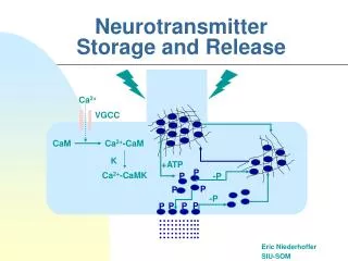
PROCESS OF TRANSMISSION STEPS : • Arrival of an action potential opens voltage-gated Ca2+ channels cytosolic Ca2+ levels • The rise in Ca2+ triggers exocytosis of the synaptic vesicles and release of NTs • The NTs diffuse across the synaptic cleft, bind to receptors on the postsynaptic membrane change membrane potential • Synaptic vesicles are endocytosed and recycled
PROCESS OF TRANSMISSION • Several inactivation mechanisms terminate the process : - enzymatic degradation of NTs - reuptake of NTs by presynaptic neuron - diffusion of NTs from the synaptic cleft • Signaling by most of the classic NTs is terminated by reuptake • Signaling by acetylcholine and neuropeptides is terminated by enzymatic degradation
SYNTHESIS OF NTs • Nonpeptide NTs are synthesized in the cytosol of axon terminals • Monoamine NTs are synthesized in a series of enzymatic steps from the precursor amino acids packaged into storage granules (synaptic vesicles).
STORAGE OF NTs • Small molecules NTs are imported from the cytosol into synaptic vesicles by a proton-coupled antiporters in the vesicle membrane and stored • Synaptic vesicles : 40-50 nm in diameter • Synaptic vesicles membrane contains V-type ATPase (proton pumps) which maintains low intravesicular pH • Synaptic vesicles membrane consist of at least eight types of membrane proteins that function in vesicle docking and fusion
RELEASE OF NTs • On stimulation of the nerve cells, NTs are released by exocytosis • Exocytosis involves vesicle - targeting and fusion • Synaptic vesicle fuse with axonal membrane releasing their contents into the synaptic cleft • Synaptic vesicle are recycled locally to the axon terminus after fusion with the plasma membrane.
Cocaine : inhibits the transporters for norepinephrine, serotonin, and dopamine. Binding of cocaine to dopamine transporter inhibits reuptake of dopamine prolonging signaling at key brain synapses. Dopamine transporter : principal brain “cocaine receptor”. Antidepressant drugs : • fluoxetine (prozac) & imipramine : block serotonin uptake • tricyclic desipramine blocks norepinephrine uptake
Proteins of the synaptic vesicle membrane • Synapsin:- a fibrous P-protein that links synaptic vesicles - phosphorylated by c-AMP-dependent protein kinase and Ca-calmodulin (CaM) kinase regulates number of free or bound vesicles - bind to cytoskeletal proteins actin and spectrin • VAMP (vesicle- associated membrane protein, synaptobrevin) : - involved in vesicle transport and exocytosis • Rab 3 (GTP-binding proteins) : - involved in docking & fusion of exocytosis. • Synaptotagmin : - contains four Ca2+ binding sites - Ca2+-sensing protein triggers vesicle exocytosis
VAMP mechanism of action of botulinum-B toxin Botulinum –B toxin : - a bacterial protein - composed of 2 polypeptides : * one peptide binds to motor neuron that release acetylcholine at neuromuscular synapse * the other, a protease, enter into the cytosol and destroy VAMP prevents acetylcholine release causing paralysis
RECEPTORS OF NEUROTRANSMITTER Two classes of NT receptors : • Ligand-gated ion channels receptors • mediate rapid postsynaptic responses (msec) • contain 5 subunits, each has a transmembrane M2 α- helix that lines the channel • NT binding triggers a conformational change leading to channel opening and permit ion passage • G-protein coupled receptors • linked to a separate ion channel regulate ion channel indirectly • mediate slow postsynaptic responses (seconds or more)
RECEPTORS OF NEUROTRANSMITTER • Depend on the specific R, the same NT can induce either excitatory or inhibitory response • Stimulation of excitatory Rs causes depolarization of postsynaptic membrane generates an action potential • Stimulation of inhibitory Rs causes hyperpolarization of postsynaptic membrane represses an action potential
ACETYLCHOLINE • Cholinergic neurons: - projection neurons : 2 clusters - basal forebrain complex - mesopontine complex • interneurons : - several brain regions including the striatum • Periphery : - preganglionic autonomic neurons - postganglionic parasympathetic neurons - neuromuscular junction
ACETYLCHOLINE • Cholinergic pathway in the brain : memory formation. • Alzheimer’s disease : majority of nucleus basalis neurons in the basal forebrain are lost leading to impairments in the cortical cholinergic innervation correlate with the severity of dementia • Dementia in Parkinson’s disease : due to degenerative process in the basal ganglia and other parts of the brain
ACETYLCHOLINE • SYNTHESIS, STORAGE AND RELEASE : • Synthesis : transfer of acetyl group from acetyl CoA to choline, catalyzed by choline acetyltransferase • Choline is derived from diet, transported from blood by high affinity transport mechanism. Choline availability is a rate limiting factor • Stored into synaptic vesicles through vesicular H+-acetylcholine antiporter • Released from the vesicles into the synaptic cleft, reacts with the nicotinic-acetylcholine receptor on the postsynaptic membrane
ACETYLCHOLINE • The action of acetylcholine is terminated by acetylcholinesterase which hydrolyzes acetylcholine to choline and acetate. Choline is transported back into the nerve terminal by a H+-choline symporter reuse Acetate is reabsorbed into the blood. • Acetylcholinesterase inhibitors is used to treat dementia of Alzheimer (augment cholinergic transmission) • Neurotoxins and nerve gasses inhibit acetylcholinesterase prolong the action of acetylcholine extending the period of membrane depolarization. Such inhibitors can be lethal if they prevent relaxation of the respiratory muscles
ACETYLCHOLINE RECEPTORS • Two major classes : - Muscarinic receptors : G-protein coupled - Nicotinic acetylcholine receptors : ligand-gated ion channels • Muscarinic : - implicated in learning & memory, sleep regulation, pain perception and regulation of seizure susceptibility - 5 subtypes which are heterogenous - M1, M3, M5 : stimulate phosphoinositides - M2 & M4 : inhibit adenylate cyclase - M1 : implicated in learning & memory processes - M4 : putative targets for anticholinergics used as antiparkinson
ACETYLCHOLINE RECEPTORS • Muscarinic : - in peripher : - M2 regulate heart rate & contractility - M3 mediate smooth muscle contraction & glandular secretion - binding of acetylcholine to muscarinic Rs in heart muscle causes dissociation of G protein open K+ channels. Influx of K+ ions hyperpolarizes the cell membrane slowing heart contraction (fig. page )
ACETYLCHOLINE RECEPTORS • Nicotinic acetylcholine Receptor : NAChR • In the brain, NAChR are found at highest densities in the area implicated in cognitive function : hippocampus, neocortex, substantia nigra, basal forebrain • Admits both K+ & Na+ • Composed of pentameric protein radially arranged around a central ion pore : subunits α2βγδ which are heterogenous • The channel opens when R cooperatively binds two ACh molecules at the interfaces of the αδ and αγ subunits • Its role is best known in synapses between motor neurons and skeletal muscle cells neuromuscular junction
ACETYLCHOLINE RECEPTORS • NAChR • Binding of ACh to NAChR at neuromuscular junction triggers a rapid increase in permiability of the membrane to Na+ & K+ ions, depolarization action potential contraction of the muscle • Cortical NAChR : - diminished in Alzheimer’s, Nicotine administration : -improves attention defects in some Alzheimer patients - improves measures of sensory gating in some schizophrenia - Some rare familial epilepsy syndromes are associated with mutation of NAChR
SEROTONIN • Serotonin systems influence CNS activity at all levels of neuraxis • Serotonergic neurons are clustered in midbrain, pons and medulla, project extensively throughout the brain and descend to the spinal cord • Two types of fibres: • Fine with small varicosities : dorsal raphe axons • Beaded with large spherical varicosities : median raphe axons • Both type of fibres are found in the neocortex, which receive serotonergic innervation from both nuclei • Caudal raphe serotonergic neurons project to medulla, cerebellum and spinal cord • MDMA (methylene-dioxy-methamphetamine, ecstasy) produces selective loss of fine axons
SEROTONIN • SYNTHESIS : • Serotonin is synthesized from tryptophan. • Brain uptake of tryptophan (active carrier mechanism) is determined by circulatory tryptophan & by ratio of tryptophan to other large neutral amino acids • Steps : - hydroxylation of tryp by tryp hydroxylase 5-hydroxytryp (rate limiting) - decarboxylation of 5-hydroxytryp by aromatic amino acid decarboxylase 5-hydroxytryptamine (serotonin)
SEROTONIN • DEGRADATION : - Mediated by monoamine oxidase type A (MAOA) which oxidizes amino group to form aldehyde - Further oxidation by aldehyde dehydrogenase 5-hydroxy- indolacetic acid (5-HIAA) • Kidney, liver tissue, fecal bacteria : convert tryptophan to tryptamine indole 3-acetate. Urinary catabolites : 5-HIAA & indole 3-acetate - MAO inhibitors : antidepressant effect elevation of serotonin
SEROTONIN RECEPTOR • Great diversity : a single NT produce a wide variety of cellular effects in multiple neuronal systems • Two types : - G protein-coupled : cAMP & inositol-3P as 2nd messengers - ligand-gated ion channels • G-protein-coupled • 5-HT1 : - the largest subfamily with subtypes - inhibits adenylate cyclase ↓ cAMP - 5-HT1A : postsynaptic & presynaptic(autoR) Stimulation of autoR suppresses activity of serotonergic neuron
SEROTONIN RECEPTOR • 5-HT2 : - stimulates phosphoinositide turnover IP3 - antidepressant, antipsychotic : antagonize 5-HT2C R - hallucinogen (LSD) : agonist activity at 5-HT2 R • 5-HT4, 5-HT6, 5-HT7 : stimulate adenylate cyclase cAMP indirectly modulate K+ channel • Ligand-gated ion channel • 5-HT3: - passage Na+ & K+ rapid excitatory effects in postsynaptic neurons
CATECHOLAMINES • DOPAMINE • Dopamine neurons : more widely distributed • Three dopamine systems : 1. nigrostriatal 2. mesocorticolimbic 3. tuberohypophyseal • Nigrostriatal: - cell bodies : in the pars compacta substantia nigra - ascending projection to dorsal striatum : modulate motor function - motor disturbances in Parkinson’s disease : degenerative disorder of nigrostriatal system - extrapyramidal adverse effects of antipsychotic drugs result from blockade of striatal receptors
DOPAMINE • 2. Mesocorticolimbic: - Ascending projection innervating limbic structures & associated cortical structures - Regulate a wide variety of stimuli, including drugs of abuse - Target of antipsychotic drugs (dopamine R antagonist) • 3. Tuberohypophyseal : - dopaminergic neurons in hypothalamic arcuate & periventricular nuclei. - projections to pituitary : inhibitory regulation of prolactin release Administration of antipsychotic drugs → galactorhea
NOREPINEPHRINE & EPINEPHRINE • Function as both systemic hormones & NTs • NE : at synapses in CNS and peripheral neurons (synapses with smooth muscles innervated by sympathetic motor neurons) • SYNTHESIS • In adrenal medulla & the brain • Hydroxylation of tyrosine to dopa by tyrosine hydroxylase • Decarboxylation of dopa to dopamine by dopa decarboxylase • Hydroxylation of dopamine to NE by dopamine β-hydroxylase, in catecholaminergic vesicles within adrenergic and noradrenergic neurons • Conversion of NE to E by phenylethanolamin-N-methyl transferase (PNMT) in adrenergic neurons
CATECHOLAMINES • DEGRADATION • The action of CA is terminated by reuptake into the presynaptic neuron repackaged into synaptic vesicles or metabolized • Metabolism by : - catechol-O-methyl transferase (COMT) & - monoamine oxidase (MAO) • COMT: catalyzes transfer of a methyl group from S-adenosyl- methionine to a phenolic -OH group • MAO : catalyzes oxidative deamination of amines to aldehydes • End product - dopamine : homovanillic acid (HVA) - NE & E : 3-methoxy-4-hydroxymandelic acid (MHMA) • CA metabolites : indicators of the activity of catecholaminergic systems
COMT : - distributed throughout the brain & peripheral tissues - wide substrate specifity - catalyzing transfer of methyl groups from S-adenosyl- methionine to hydroxyl group of catechol compouds • MAO : - located on the outer membrane of mitochondria - oxidatively deaminates catecholamines to aldehydes - two isoenzymes : MAOA & MAOB - MAOA: preferentially deaminates serotonin & NE - MAOB: deaminates histamine, dopamine & phenylethylamine - in peripheral tissues (GI & liver) : prevent accumulation of toxic amines • MAO inhibitor : - block MA catabolism monoamines in the brain - adverse effects : peripheral amines
CATECHOLAMINES RECEPTORS • All known CA Rs are coupled to G proteins • Different Rs are linked to different G pr different 2nd messengers • DOPAMINE Rs : D1-5 • D1 : - stimulates - adenylate cyclase cAMP - phosphoinositide turnover IP3 - not found on dopaminergic neuron ( not an autoR) - contribute to the effects of cocain in CNS - low affinity for antipsychotic (butyrophenones, halloperidol) • D5 , D1-like : - structural similarity - stimulates adenylate cyclase
DOPAMINE RECEPTOR • D2 : - interacts with a variety of G pr diverse 2nd messenger - ↓ cAMP - modulates Ca2+ & K+ channel - alters phosphoinositide production - functions as postsynaptic or autoR on dopaminergic terminals, cell bodies & dendrites of dopaminergic neuron - high affinity for antipsychotic drugs - in ant. pituitary : inhibition of prolactin & MSH release - in schizophrenia : D2R - extrapyr adverse effects of antipsychotic: block of striatal D2R • D3,D4 , D2-like :- similar in structure & pharmacology - D4R : more abundant in heart than in the brain - in schizophrenia : D4R