Download
1 / 88
930 likes | 1.17k Views
The Organic Chemistry of Drug Design and Drug Action. Chapter 6 DNA-Interactive Agents. DNA-Interactive Agents. DNA - another receptor Carries genetic information in cells Few differences between normal DNA and DNA from other cells.

E N D
The Organic Chemistry of Drug Design and Drug Action Chapter 6 DNA-Interactive Agents
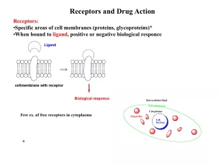
DNA-Interactive Agents DNA - another receptor Carries genetic information in cells Few differences between normal DNA and DNA from other cells. Therefore, these drugs are generally very toxic; used for life-threatening diseases, such as cancer and viral infections.
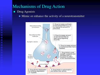
Cancer Cells Rapid, abnormal cell division Constant need for DNA and precursors • Selective toxicity • rapid uptake of drug molecules by cancer cells • repair mechanisms too slow • activation of proteins such as p53 in normal cells in response to DNA damage - leads to increased DNA repair enzymes, cell cycle arrest (to allow time for DNA repair), and programmed cell death (apoptosis)
Structure-Based Design of Potential Drugs for Prevention of Hair Loss During Chemotherapy Inhibition of cyclin-dependent kinase 2 (CDK2) arrests hair follicle cell cycle, rendering it less susceptible to anticancer agents. Lead: Known inhibitors of tyrosine kinases
Lead Modification Inhibits CDK2 with IC50 60 nM
Crystal Structure of 6.2 and 6.3 Bound to CDK2 Figure 6.1 Note that the inhibitor is near Lys-33 (need a H bond acceptor) and Val-18 (need a hydrophobic group); the SO2NH2 group can be substituted. Note the N of the thiazole can H bond to Lys-33, and the S of the thiazole is hydrophobic. The pyridine does not interfere, but can increase solubility.
Combination Chemotherapy • In the late 1950s combination chemotherapy was introduced. • Effectiveness compared to single drug: • Able to fight acquired resistance • Different mechanisms of action increase effectiveness • Some covalent modifications can be reversed by repair enzymes, so inhibitors of DNA repair can be added
Drug Interactions Care must be given to which mechanisms of action are involved in drug combinations. For example, a renal (kidney) cytotoxic agent should not be used with a drug that requires renal elimination for excretion.
Drug Resistance 1. Increased expression of membrane glycoproteins - affects membrane permeability (blocks drug transport) 2. Increased levels of thiols (destroys electrophilic anticancer drugs) 3. Increased levels of deactivating enzymes (destroys anticancer drugs) 4. Decreased levels of prodrug-activating enzymes (prevents activation of prodrugs) 5. Increased DNA repair enzymes (repairs DNA modification) All involve gene alterations.
DNA Structure and Properties purine adenine pyrimidine cytosine purine guanine pyrimidine thymine In double-stranded DNA the ratio of A/T and G/C is always 1.
Hydrogen Bonding of Complementary Base Pairs (Watson-Crick Base Pair) 2 H-bonds
Hydrogen Bonding 3 H-bonds
The 2 glycosidic bonds that connect the base to its sugar are not directly opposite each other, giving different spacings along helix. Figure 6.3
Duplex (double-stranded) DNA Figure 6.4 (all inside)
Figure 6.5 most stable tautomer
Figure 6.6 mimics thymine mimics adenine These can substitute for T and A in DNA polymerase reactions. Therefore H bonding is not essential; only need the groups to fit snugly in the binding site of DNA polymerase.
DNA Shapes Human somatic cells - each of the 46 chromosomes consists of a single DNA duplex about 4 cm long. Therefore a total of 46 4 = 1.84 m long of DNA packed into the nucleus. Nucleus is only 5 m in diameter Done with aid of richly basic proteins called histones. Folded compact form of DNA called chromatin.
Packing of DNA into the Nucleus Figure 6.7
Supercoiled DNA - Packing of Bacterial DNA Facilitates RNA polymerase reaction Helps in chromatin packing Figure 6.9 circular DNA (plasmid) supercoiled DNA Enzymes that interconvert supercoiled and relaxed DNA are called DNA topoisomerases.
DNA topoisomerases also resolve topological problems such as catenation and knotting. Figure 6.10 catenanes
Figure 6.11 knots
Two Principal Types of Topoisomerases DNA topoisomerases I catalyze transient breaks of one strand of duplex DNA. DNA topoisomerases II (in bacteria called DNA gyrase) catalyze cleavage of both strands of duplex DNA.
Mechanisms of Topoisomerase IA and IB Scheme 6.1
Possible Mechanism of Topoisomerase I Reaction Conformational change to make a gap for strand to pass through Attack of Tyr at 5-phosphate Cleavable complex Religation of the two ends Ready for another catalytic cycle Relaxed DNA is released Figure 6.12
Mechanism for Topoisomerase I Decatenation (B) Figure 6.13
DNA Conformations Figure 6.14 Right-handed helices Left-handed helix
A- and B-DNA glycosyl bonds are always anti. anti (base in the opposite direction as the 5-phosphate)
Z-DNA glycosyl bond is anti at pyrimidines but syn at purines (responsible for zigzag appearance). syn (base in the same direction as the 5-phosphate)
Classes of DNA-Interactive Drugs Reversible binders - reversible interactions with DNA Alkylators - react covalently with DNA bases Strand breakers - generate reactive radicals that cleave polynucleotide strands
How Do Drugs Interact with DNA Packed as Chromatin? Figure 6.15A Figure 6.15B The outer surface of the DNA is accessible to small molecules.
Also, nucleosomes are in dynamic equilibrium with uncoiled DNA, so drug can bind after uncoiling. Figure 6.16
Reversible DNA Binders Three ways small molecules can reversibly bind to duplex DNA. External electrostatic Groove binder Intercalation Figure 6.17
External electrostatic binders - cations that bind to anionic phosphates. Groove binders - proteins prefer major groove binding; small molecules prefer minor groove binding. Minor groove generally not as wide in A-T regions as in G-C regions. Therefore, flat aromatic, often crescent-shaped molecules (6.11) prefer A-T regions.
DNA Intercalators Flat, generally aromatic or heteroaromatic molecules Insert (intercalate) and stack between base pairs Noncovalent interactions Drug is perpendicular to helix axis Sugar-phosphate backbone is distorted Energetically favorable process Does not disrupt H-bonding Destroys regular helix; unwinds DNA Therefore interferes with the action of DNA topoisomerases and DNA polymerases, which elongate DNA chain and correct mistakes in the DNA
Example of Intercalation Figure 6.18
Topotecan binds to the DNA-topoisomerase I complex antitumor agent Does not appear to be a correlation between DNA intercalation and antitumor activity. It is not sufficient to intercalate without stabilization of the cleavable complex.
Selected Examples of DNA Intercalators Acridines Actinomycins Anthracyclines
Amsacrine - acridine analog Lead modification Lead compound Proflavine antibacterial anti-leukemia agent stabilizes cleavable complex
Crystal Stucture of an Actinomycin Analog Bound to a DNA Figure 6.19 dactinomycin - antitumor from Streptomyces Resistance - efflux pump (P170 glycoprotein) and impaired drug uptake
Anthracycline Analog Figure 6.20 Complex stabilized by stacking energy and H-bonding D ring (major groove) A ring (minor groove) daunorubicin (daunomycin) Intercalation and topoisomerase II-induced damage anti-leukemia agent
DNA Alkylators Nitrogen mustards Lead discovery Autopsies of soldiers killed in World War I by sulfur mustard (6.23) showed leukopenia (low white blood cells), bone marrow defects, dissolution of lymphoid tissue, ulceration of GI tract. These are all rapidly replicating cells. sulfur mustard Suggested this may show tumor cytotoxicity too. 1931 - S mustard tried as antitumor agent, but too toxic.
Lead Modification Less toxic form of sulfur mustard sought. 1942 - first clinical trials of a nitrogen mustard Marks beginning of modern cancer chemotherapy (for advanced Hodgkin’s disease)
Chemistry of Alkylating Agents Scheme 6.2 Reactivity of Nu- in general: RS- > RNH2 > ROPO3= > RCOO-
For DNA: N-7 of guanine > N-3 of adenine > N-7 of adenine > N-3 of guanine > N-1 of adenine > N-1 of cytosine N-3 of cytosine, the O-6 of guanine, and phosphate groups also can be alkylated. Purines A/G Pyrimidines T/C
Scheme 6.3 anchimeric assistance If k1 > k2, SN2 If k2 > k1, SN1 • Bifunctional alkylating agents • DNA undergoes intrastrand and interstand cross-linking • Compounds that cross-link DNA (bifunctional alkylating agents) are much more effective.
Hydrolysis of alkylated N-7 guanine leads to destruction of the purine nucleus. Scheme 6.4
Mechlorethamine is quite unstable to hydrolysis (completely reacts within minutes of injection). Therefore, a more stable analog is needed. 6.29 More stable Slows rate of aziridinium formation R = COOH too stable, but soluble R = (CH2)3COOH chlorambucil
Ethylenimines Lower pKa of the aziridine N so it is not protonated at physiological pH - attach e--withdrawing group Need at least 2 aziridines per molecule for antitumor activity e--withdrawing group