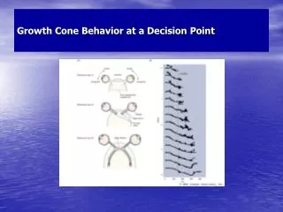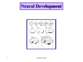
Neural development (growth cones)
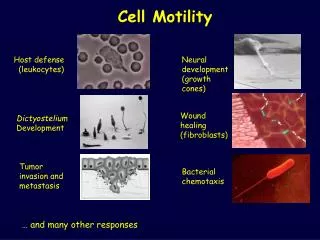
Cell Motility. Host defense (leukocytes). Neural development (growth cones). Wound healing (fibroblasts). Dictyostelium Development. Tumor invasion and metastasis. Bacterial chemotaxis. … and many other responses. Amoeboid Chemotaxis.

Neural development (growth cones)
E N D
Presentation Transcript
Cell Motility Host defense (leukocytes) Neural development (growth cones) Wound healing (fibroblasts) Dictyostelium Development Tumor invasion and metastasis Bacterial chemotaxis … and many other responses
Amoeboid Chemotaxis Mesenchymal cells: stem cells that can differentiate into fibroblasts, fat cells, mast cells and osteoblasts, etc. (original definition)
Amoeboid Chemotaxis Human neutrophils Gold fish fibroblast Green: GFP-actin; Red: rhodamine-vinculin Vinculin: focal adhesion marker
Mesenchymal cells: stem cells that can differentiate into fibroblasts, fat cells, mast cells and osteoblasts, etc. (original definition)
A mouse embryonic fibroblast Gold fish fibroblast DSRed-paxillin Paxillin; a focal adhesion marker Webb et al., Nat Cell Biol 4, E97 - E100 (2002)
Dynamics of GFP-Zyxin and Traction Forces during Cell Migration Traction-induced deformation of substrate (red: “hot” spot) Zyxin: another focal adhesion marker Fish fibroblast Nascent focal adhesions generally exert stronger force ! Beningo et al., J Cell Biol. 2001 153:881
GST-bni1p Maximal nucleation
Processive movement Higashida et al., Science, 2004, 303:2007
Rho-GTP activates ROCK, which in turn activates myosin ROCK (a serine-threonine kinase) activates MLC directly (ser 19 phosphorylation) and also inhibits MBS (through phosphorylation) (phosphorylation of myosin high ATPase activity) Autoinhibition again ! Nature Reviews Mol Cell Biol, 2003, 4, 446 MLC: myosin light chain MBS: myosin binding subunit (myosin phosphotase)
Mesenchymal Migration: is this really physiologically relevant ?
Tissue microenvironment in vivo Epithelial cells fibroblasts
Cell culture in 3-dimension BM: basement membrane (extracellular matrix) Weaver et al., J Cell Biol. 1997 137:231 Petersen et al., Proc Natl Acad Sci U S A. 1992 89:9064
Cell Migration in 3D In 3D collagen and in granulation tissues in vivo, fibroblast migration occurs in the absence of actin stress fiber formation, .. and instead exhibits a diffuse cortical F-actin distribution over the entire cell body Welsh et al., J Cell Biol, 1990, 110, 133 Grinnell, J Cell Biol, 1994, 124, 4, 401
Tumor invasion in vitro and in vivo involves the proteolytic degradation of ECM barriers • However, after blocking of MMPs or serine proteases, significant residual migration of individual cells is observed in different migration models • In vivo, protease inhibitor–based targeting of MMPs and serine proteases has yielded an unexpectedly weak benefit in some animal tumor models as well as clinical trials in humans, suggesting that a principal dissemination capacity remained intact What has happened? Tumor Cell migration in 3-D (“Supramolecular plasticity”)
Tumor Cell migration in 3-D HT1080/MT1-MMP fibrosarcoma cells (Fibroblast-like, mesenchymal cells) 3D collagen matrix MMP: matrix metalloprotease Matrix defect Wolf et al., J Cell Biol, 160, 2003 267–277
Proteases-independent cell migration in 3D Matrix fiber breakdown FITC release J Cell Biol, 160, 2003 267–277
Transition of mesenchymal to amoeboid movement in the presence of protease inhibitors J Cell Biol, 160, 2003 267–277
In vivo translocation and morphology of cells in mouse dermis Co-injection of control HT1080 cells (green) and cells pretreated with protease inhibitors (red) Injection site J Cell Biol, 160, 2003 267–277
Supramolecular plasticity mechanism in (tumor) cell migration J Cell Biol, 160, 2003 267–277
Comments/Questions: • Does the mesenchymal to amoeboid transition occur • on 2D? • 2. Amoeboid, really? • 3. Concerns about co-injection experiment • 4. The signaling mechanisms
The signaling mechanisms Tumor cells Sahai et al., 2003, Nat Cell Biol, 5, 711
Rho or ROCK function and Cell Morphology C3 toxin: Rho inhibitor Y 27632: ROCK inhibitor F-actin Cortactin 3D matrix Sahai et al., 2003, Nat Cell Biol, 5, 711
Different modes of motility has different requirements for RhoA and ROCK signaling RhoA (G14V) ROCKΔ3 Rac (T17N) DN mutant C3 toxin Rho inhibitor Y 27632 ROCK inhibitor DP mutants F-actin staining (co-transfection: EGFP + mutants) Classic epistasis experiment! Rho activity is required for round morphology; Rac activity is required for elongated morphology Sahai et al., 2003, Nat Cell Biol, 5, 711
Rounded Cell Motility, 3D vs. 2D A375 m2 cells on 2D plastic Chemoattractants (serum growth factors) Direction of Cell migration 3D Matrix A375 m2 cells on 2D plastic +Y 27632 A375 m2 cells (2- 5 mm/hour) Sahai et al., 2003, Nat Cell Biol, 5, 711
Combined treatment of tumor cells with Protease Inhibitors and Y 27632 Tumor cell invasion into ECM Rounded motility does not require pericellular proteolysis PI has minimal effects Combined treatment has strong effects Sahai et al., 2003, Nat Cell Biol, 5, 711