Download
1 / 43
430 likes | 1.15k Views
Cerebral Palsy The ABC’s of CP. Toni Benton, M.D. Continuum of Care Project UNM HSC School of Medicine April 20, 2006. Cerebral Palsy. Outline I. Definition II. Incidence, Epidemiology and Distribution III. Etiology IV. Types V. Medical Management VI. Psychosocial Issues VII. Aging.

E N D
Cerebral PalsyThe ABC’s of CP Toni Benton, M.D. Continuum of Care Project UNM HSC School of Medicine April 20, 2006
Cerebral Palsy Outline I. Definition II. Incidence, Epidemiology and Distribution III. Etiology IV. Types V. Medical Management VI. Psychosocial Issues VII. Aging
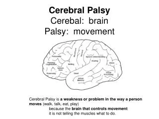
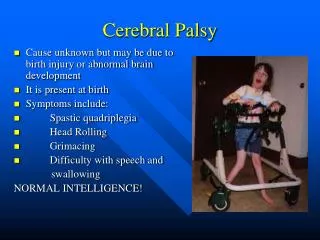
Cerebral Palsy-Definition Cerebral palsy is a symptom complex, (not a disease) that has multiple etiologies. CP is a disorder of tone, posture or movement due to a lesion in the developing brain. Lesion results in paralysis, weakness, incoordination or abnormal movement Not contagious, no cure. It is static, but it symptoms may change with maturation
Cerebral Palsy Brain damage Occurs during developmental period Motor dysfunction Not Curable Non-progressive (static) Any regression or deterioration of motor or intellectual skills should prompt a search for a degenerative disease Therapy can help improve function
Cerebral Palsy • There are 2 major types of CP, depending on location of lesions: • Pyramidal (Spastic) • Extrapyramidal • There is overlap of both symptoms and anatomic lesions.
The pyramidal system carries the signal for muscle contraction. • The extrapyramidal system provides regulatory influences on that contraction.
Cerebral Palsy • Types of brain damage • Bleeding • Brain malformation • Trauma to brain • Lack of oxygen • Infection • Toxins • Unknown
Epidemiology • The overall prevalence of cerebral palsy ranges from 1.5 to 2.5 per 1000 live births. • The overall prevalence of CP has remained stable since the 1960’s. • Speculations that the increased survival of the VLBW preemies would cause a rise in the prevalence of CP have proven wrong. • Likewise the expected decrease in CP as a result of C-section and fetal monitoring has not happened. • However, the prevalence of the subtypes has changed.
Epidemiology • Due to the increased survival of very low birth weight preemies, the incidence of spastic diplegia has increased. • Choreoathetoid CP, due to kernicterus, has decreased. • Multiple gestation carries an increased risk of CP.
Etiology • CP has multiple etiologies- many are still unknown • Since CP is not a single entity, recurrence risks depend on the underlying cause. • If there is a regression in skills, suspect a degenerative disease.
Etiology • Most causes are prenatal- genetic, congenital malformations, metabolic, intrauterine infections, rather than perinatal or postnatal- birth asphyxia, hemorrhage, infarction, infections, trauma.
Etiology • Much of the literature of the 1990’s was directed at the controversy re the role of asphyxia in the etiology of CP • Asphyxia implies poor gas exchange, low Apgars and neurologic depression during and soon after delivery. • Significant asphyxia is accompanied by acidosis. • Asphyxia is rarely the cause of CP in the term infant.
Etiology • In one outcome study of 43,437 full term children, 150 had cerebral palsy. Only 9 of these cases were attributable to birth asphyxia. • 34 had spastic quadriplegia and 71% of those cases had identifiable causes. 53%- congenital disorders 14%-birth asphyxia 8%-CNS infections
Etiology • Among the children with non quadriplegic cerebral palsy, congenital disorders appeared to account for about 1/3 of the cases, and CNS infections accounted for 5%. (Wilson and Cooley-2000; Collaborative Perinatal Study of The National Institute of Neurological and Communicative Disorders and Stroke,Naeye, 1989)
Hypoxic Ischemic Encephalopathy (HIE) • A clinical entity first described in 1976 • Used interchangeably with Neonatal encephalopathy. • Asphyxia refers to the first minutes after birth (low Apgars and acidosis) • HIE signs and symptoms persist over hours and days that follow.
Hypoxic Ischemic Encephalopathy (HIE) 3 major lesions arise from HIE • Periventricular Leukomalacia (PVL) Typically seen in the premature infant a. Hemorrhagic PVL b. Ischemic PVL • Parasaggital Cerebral Injury Typically seen in the term infant • Selective (Focal) Neuronal Necrosis Seen in both term and premature infants
Periventricular Leukomalacia (PVL) • Hemorrhagic PVL • Hemorrhage is associated with a collection of primitive cells between the ependyma and caudate that are programmed to “melt away” at 32-34 weeks gestation • They contain fragile capillaries that are easily damaged by hypoxia (lack of oxygen) and hypotension (drop in blood pressure). • When the blood pressure returns to normal, bleeding occurs because the preemie has underdeveloped autoregulation.
Periventricular Leukomalacia (PVL) • Hemorrhagic PVL(cont.) • This bleeding may then rupture into the ventricle and/or parenchyma • Periventricular venous congestion (swelling) may then occur, and cause ischemia (lack of blood supply) and periventricular hemorrhagic infarction.
Periventricular Leukomalacia (PVL) 2. Ischemic PVL • An ischemic infarction or failure of perfusion usually to the watershed area surrounding the ventricular horns- “HIE white matter necrosis”. • Peak incidence occurs around 32 weeks • Larger infarcts may leave a cyst • Secondary hemorrhage can occur into theses cysts- “periventricular hemorrhage”.
Periventricular Leukomalacia (PVL) 2. Ischemic PVL • PVL can extend into the internal capsule and result in hemiplegia superimposed on diplegia. • Prenatal maternal ultrasound has detected lesions in the fetus at 28-32 weeks gestation, thus confirming that PVL can occur prenatally.
Parasaggital Cerebral Injury • Injury is related to vascular factors, especially in the parasaggital border zones that are more vulnerable to a drop in perfusion pressure and immature autoregulation. • The ischemic lesion results in cortical and subcortical white matter injury. • It is usually bilateral and symmetric. • The posterior aspect of the cerebral hemisphere especially the parietal occipital regions is more affected than the anterior.
Selective (Focal) Neuronal Necrosis (SNN) • Occurs in the glutamate sensitive areas in the basal ganglia, thalamus, brainstem and cortex. • The location of the focal necrosis, which show up as cystic lesions on MRI, depend on the stage of development of the infant’s brain at the time of the HIE. • For example, HIE at term often produces SNN in the basal ganglia since it is glutamate sensitive and very hypermetabolic at term.
Pyramidal Described as a Clasped knife response or Velocity dependent increased resistance to passive muscle stretch The spasticity can be worse when the person is anxious or ill. The spasticity does not go away when the person is asleep. Extrapyramidal Ataxia Hypotonia Dystonia Rigidity The tone may increase with volitional movement, or when the person is anxious During sleep the person is actually hypotonic Types of Cerebral Palsy
Types of Cerebral Palsy • Pyramidal (Spastic) • Quadriplegia- all 4 extremities • Hemiplegia- one side of the body • Diplegia- legs worse than arms • Paraplegia- legs only • Monoplegia- one extremity
Dyskinetic Athetosis- slow writhing, wormlike Chorea- quick, jerky movements Choreoathetosis- mixed Hypotonia- floppy, low muscle tone, little movement Ataxic CP Results from damage to the cerebellum Ataxia- tremor & drunken- like gait B. ExtrapyramidalDivided into Dyskinetic and Ataxic types
Pyramidal Lesion is usually in the motor cortex, internal capsule and/or cortical spinal tracts. Extrapyramidal Lesion is usually in the basal ganglia, Thalamus, Subthalamic nucleus and/or cerebellum. Anatomy
Medical Management Growth • Persons with CP often have struggle to gain or maintain weight. • Failure to Thrive is a common problem. • Before diagnosing Failure to thrive, an accurate Body Mass Index must be obtained, but an accurate height is difficult to obtain in a person with severe contractures. • In such cases, arm span calculations may be used and a growth chart is available to determine percentiles standardized to age and gender.
Medical Management Orthopedic Problems • Scoliosis • Hip Dislocations • Contractures • Osteoporosis
Medical Management Oromotor Dysfunction • Especially common in persons with Extrapyramidal CP and Spastic quadriplegia • Language delay/Speech delays • Drooling • Dysphagia • Aspiration
Medical Management Gastrointestinal Dysmotility • Delayed gastric emptying • Gastroesophageal reflux • Pain • Chronic aspiration • Constipation These disorders are interrelated and compound one another.
Medical Management Spasticity Management Management of spasticity does not fix the underlying pathology of CP, but it may decreased the sequelae of increased tone. • Over time, the spasticity leads to: • musculoskeletal deformity • scoliosis • hip dislocation • contractures • Pain • Hygiene problems
Treatment of Spasticity Medications • Valium • Dantrium • Baclofen • Clonidine • Clonazepam • BOTOX
Treatment of Dystonia Medications-(None are very effective) • L-Dopa- drug of choice for certain disorders • Artane • Anticonvulsants-for intermittent and paroxysmal dystonia • Anti-spasticity medications- • Haldol or Reserpine- for choreoathetosis • Propranolol- for essential tremor • Clonazepam or Valium- for “rubral tremors”-(course tremors of the entire arm) • Valproic acid or clonazepam for action myoclonus- (large jerks with intentional movements)
Mental Retardation Communication Disorders Neurobehavioral Seizures Vision Disorders Hearing loss Somatosensation (skin sensation, body awareness) Temperature instability Nutrition Drooling Dentition problems Neurogenic bladder Neurogenic bowel Gastroesophageal reflux Dysphagia Autonomic dysfunction Associated Problems
Other Treatments • Casting • Therapeutic Electrical Stimulation • Patterning: Doman-Delacato- (not recommended) • Selective Dorsal Rhizotomy • Massage • Hyperbaric Oxygen • Acupuncture
Adult Concerns Medical • Routine Healthcare Maintenance • Sequelae of Spasticity • Orthopedic Issues • Pain Management • Neurogenic Bowel and Bladder • Prevention of Chronic Aspiration Management of Gastroesophageal Reflux & Complications • Barrett’s esophagus • Esophageal strictures • Esophageal/stomach cancer
Adult Concerns Psychosocial Transition from Pediatric to Adult services Independence Work Home Relationships Guardianship End of life