Download
1 / 17
180 likes | 459 Views
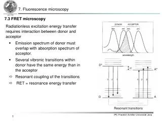
7. Fluorescence microscopy. 7.3 FRET microscopy. Radiationless excitation energy transfer r equires interaction between donor and acceptor Emission spectrum of donor must overlap with absorption spectrum of acceptor.

E N D
7. Fluorescence microscopy 7.3 FRET microscopy • Radiationless excitation energy transfer requires interaction between donor and acceptor • Emission spectrum of donor must overlap with absorption spectrum of acceptor. • Several vibronic transitions within donor have the same energy than in the acceptor • Resonant coupling of the transitions • RET = resonance energy transfer Resonant transitions
7. Fluorescence microscopy 7.3 FRET microscopy • Assumption: 2 electrons one at the donor D and one at the acceptor A are involved in the transition: • Antisymmetric wavefunction (Fermions) for initially excited state i (D excited, but not A) and final state f (A excited, but not D):Overall Hamiltonian:Interaction energy: Radiationless excitation energy transfer Coulomb term UC Exchange term Uex
7. Fluorescence microscopy 7.3 FRET microscopy Radiationless excitation energy transfer Coulomb Interaction (CI) Exchange Interaction
7. Fluorescence microscopy 7.3 FRET microscopy Radiationless excitation energy transfer • Different interaction mechanism lead to excitation energy transfer: Dipolar(Förster) „LongRange“ Coulombinteraction Multipolar Singlet energy transfer Electron exchange(Dexter) „ShortRange“ Inter molecular orbital overlap Charge resonance interaction Triplet Energy transfer
7. Fluorescence microscopy 7.3 FRET microscopy Radiationless excitation energy transfer • Coulomb interaction dominates for allowed i.e. singlet-singlet-transitions. • For forbidden transitions i.e. singlet-triplet-transitions exchange-interaction (only acting for short distances < 10 Å because overlap of orbitals is necessary) dominates. • Coulomb interactions appears also for larger distances up to 80 – 100 Å. • Interaction strength depends on interaction energy (U), energy distance between D* and A* (E), absorption bandwidth (w) and vibronic bandwidth (). Strong coupling: U>>DE U>>Dw,De Weak coupling: U>>DE Dw>>U>>De Very weak coupling: U<<De<<Dw
7. Fluorescence microscopy 7.3 FRET microscopy Förster Resonance Energy Transfer (very weak coupling): D + hn1 D* Absorption D* + A A* + D Energy transfer A* A + hn2 Emission • The following conditions must hold: • D must be a fluorophore with sufficiently long life-time • Partial spectral overlap between emission spectrum of D and absorption spectrum of A • Transition dipole moments D and A must be oriented properly to each other; • Distance between D and A shouldn‘t be too large
7. Fluorescence microscopy 7.3 FRET microscopy Förster Resonance Energy Transfer (very weak coupling): • Coulomb interaction can be developed in a multipole series in which the dipole term exhibits the term with the longest range • Energy transfer via dipole-dipole transfer has been first calculated by Förster and is therefore called Förster process • Energy transfer rate from molecule D to molecule A at a distance r: kD = radiative decay rate of donor tD0 = donor life-time in absence of energy transfer r-6-dependency as a result of dipole-dipole interaction R0 = critical distance or Förster-radius (distance at which intensity decrease caused by energy transfer and spontaneous decay are equal ( = kD)).
7. Fluorescence microscopy 7.3 FRET microscopy Förster Resonance Energy Transfer (very weak coupling): • R0 can be determined via spectroscopic values: • For R0 in Å, l in nm, eA(l) in M-1 cm-1 (overlap integral in M-1 cm-1 nm4) • Typical values for Förster-radii R0, i.e. for distances, over which energy transfer is important lie in the range of 15 -60 Å Overlap between fluorescence of donor and absorption of acceptor k2 = orientational factorF0D = quantum yield of donor in absence of energy transfer n = average refractive index for wavelength area of spectral overlapID(l) = normalized fluorescence spectrum of donor ( ) eA(l) = molar absorption coefficient of acceptor.
7. Fluorescence microscopy 7.3 FRET microscopy Förster Resonance Energy Transfer (very weak coupling): • Transfer efficiency can be expressed by: • In combination with changed lifetime: • It follows: distance dependency: D und D0 are excited state life-times of donor in absence and presence of acceptor, respectively
mD mA 7. Fluorescence microscopy 7.3 FRET microscopy Förster Resonance Energy Transfer (very weak coupling): • Besides the distance between the two chromophores also the relative orientation of the transition dipole moments of the donor D and acceptor A plays a crucial role for the energy transfer efficiency • The orientation factor k2 is given by: A: angle between D-A connecting line and acceptor transition dipole moment D: angle between D-A connecting line and donor transition dipole moment T: angle between donor and acceptor transition dipole moment
7. Fluorescence microscopy 7.3 FRET microscopy Förster Resonance Energy Transfer (very weak coupling): • For systems where the orientation stays constant during the energy transfer (e.g. usage of highly viscose solvents or rigid coupling of chromophores to large and stiff molecules) k2 can reach values between 0 (transition dipole moments are orthogonal) and 4 (collinear arrangement);k2 = 1, for a parallel arrangement • If both acceptor and donor can rotate the orientational factor 2 must be replaced by an average value: • In case both chromophores undergo a fast isotropic rotation i.e. the rotation is considerably faster than the energy transfer rate the average orientation factor is given by k2 = 2/3 • In case donor and acceptor are freely movable but the rotation is significantly slower than the energy transfer the orientation factor results in: 2 = 0.476
7. Fluorescence microscopy 7.3 FRET microscopy Förster Resonance Energy Transfer (very weak coupling): • RET is utilized as „optical nano ruler“ (10 – 100 Å) in biochemistry and cell biology • Distance between donor and acceptor should be in the range of:because R0 is a benchmark for donor-acceptor distances which can be determined by FRET.
7. Fluorescence microscopy 7.3 FRET microscopy Förster Resonance Energy Transfer (very weak coupling): • RET as „optical nano ruler“ in biochemistry and cell biology
7. Fluorescence microscopy 7.3 FRET microscopy Förster Resonance Energy Transfer (very weak coupling): • RET as „optical nano ruler“ in biochemistry and cell biology
7. Fluorescence microscopy 7.3 FRET microscopy Förster Resonance Energy Transfer (very weak coupling): • RET as „optical nano ruler“ in biochemistry and cell biologyOne requires appropriate method to label specific intracellular proteins with suitable fluorophores (fluorescent proteins genetics): • Green Fluorescent Protein (GFP) first isolated from the jellyfish Aequorea victoria GFP can be combined with just about any other protein by attaching its gene to the gene of a target protein, thereby introducing it into a cell. Thus by recording the GFP fluorescence the spatial and temporal distribution of this target protein can be directly monitored in living cells, tissue and organism. Several GFP mutants with altered fluorescence spectra exist. These mutants are named according to their color e.g. CFP (cyan) or YFP (yellow) Excitation maxima at 395 und 475 nmEmission wavelength at 509 nm
7. Fluorescence microscopy 7.3 FRET microscopy Agar plate of fluorescent bacteria colonies
7. Fluorescence microscopy 7.3 FRET microscopy Förster Resonance Energy Transfer (very weak coupling): • RET as „optical nano ruler“ in biochemistry and cell biology :GFP-mutants FRET R0 = 4.7 – 4.9 nm no FRET protein folding protein-protein interaction
![Blue-Colored Donor-Acceptor [2]Rotaxane](https://cdn1.slideserve.com/2419410/blue-colored-donor-acceptor-2-rotaxane-dt.jpg)