Download
1 / 38
380 likes | 504 Views
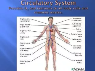
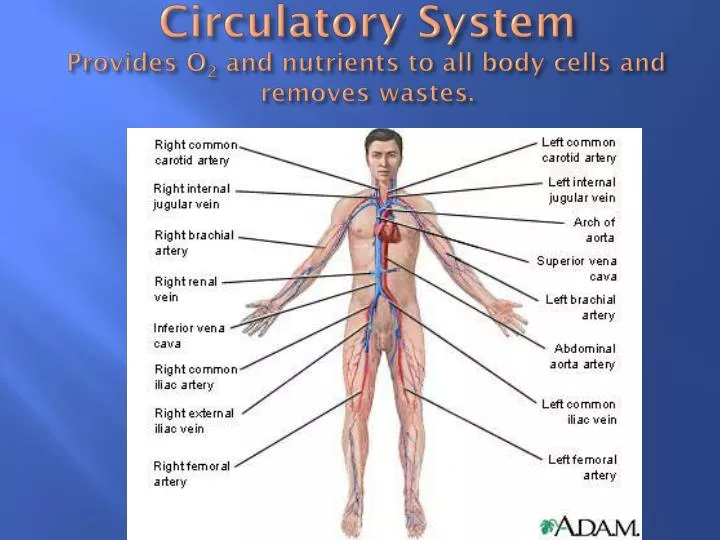
Circulatory System Provides O 2 and nutrients to all body cells and removes wastes. Structure of the Heart - Fig. 12-1, 12-2; located within mediastinum and rests on the diaphragm . Coverings of the heart Enclosed in a layered pericardium Pericardial space between layers is fluid filled

E N D
Circulatory SystemProvides O2 and nutrients to all body cells and removes wastes.
Structure of the Heart - Fig. 12-1, 12-2; located within mediastinum and rests on the diaphragm • Coverings of the heart • Enclosed in a layered pericardium • Pericardial space between layers is fluid filled • Wall of heart • Endocardium – inner layer • Myocardium – middle; mostly cardiac muscles • Epicardium (visceral pericardium) – outer layer
Structure of the Heart - Fig. 12-1, 12-2; located within mediastinum and rests on the diaphragm • Heart Chambers, Valves & Blood Flow • Heart is divided into 4 chambers – 2 atria, 2 ventricles • R. chambers and valves – O2 poor blood • R. atrium receives blood from superior & inferior vena cava & coronary sinus • Blood passes through tricuspid valve to R. ventricle • Blood passes through pulmonary semilunar valve to L. & R. pulmonary arteries ***only arteries to carry O2 poor blood***
Structure of the Heart - Fig. 12-1, 12-2; located within mediastinum and rests on the diaphragm • Heart Chambers, Valves & Blood Flow • L. chambers and valves – O 2 rich • Blood returns from lungs via L. & R. pulmonary veins ***only veins to carry O2 rich blood*** • O2 rich blood dumps into L. atrium • Blood passes through bicuspid valve into L. ventricle • Blood passes through aortic semilunar valve to aorta • Distributed to the rest of body (systemic circulation)
Structure of the Heart - Fig. 12-1, 12-2; located within mediastinum and rests on the diaphragm
Structure of the Heart - Fig. 12-1, 12-2; located within mediastinum and rests on the diaphragm
Structure of the Heart - Fig. 12-1, 12-2; located within mediastinum and rests on the diaphragm • http://www.youtube.com/watch?v=mH0QTWzU-xI (blood flow through the heart) • http://vimeo.com/8321006 (animation - blood flow)
Actions of the Heart • Cardiac Cycle • Pressure within chambers rises & falls in repeated cycles • Contraction of heart – systole Relaxation of heart – diastole • When atria are relaxes (atrial diastole) blood flows into them from veins (about 70% of blood flows directly into ventricles) • When atria contract (atrial systole) the remaining 30% of blood flows into ventricles • As ventricles contract (ventricular systole) bicuspid/tricuspid valves are pressed closed; blood flows either to lungs or body • Stroke volume = volume of blood ejected from ventricles • http://www.youtube.com/watch?v=jLTdgrhpDCg
Actions of the Heart • Heart Sounds • Described as lub-dub • Due to the vibrations produced by the blood & valve movements • Lub – occurs as A-V valves are closing/ventricles contract • Dub – occurs as semilunar valves are closing/ventricles relax
Actions of the Heart • http://www.youtube.com/watch?v=te_SY3MeWys
Actions of the Heart • Cardiac Conduction System – Fig. 12-7 • Composed of specialized cardiac muscle tissue and functions to initiate and conduct depolarization waves through the myocardium • Signal is initiated by S-A node located in upper part of R. atrium (known as the pace maker of the heart). • Ability to excite themselves • Impulses spread into surrounding myocardium • Atria contract
Actions of the Heart • Cardiac Conduction System – Fig. 12-7 • Impulses travel slowly from S-A node (so atria have time to contract) to A-V node located in lower part of R. atrium • Impulses now travel quickly to A-V bundle (bundle of His) and then to Purkinje fibers • Ventricles contract – muscle fibers in ventricular walls are arranged in whorls that “wring” blood out of ventricles
Actions of the Heart • Regulation of the Cardiac Cycle • Heartbeat is affected by physical exercise, body temp. and concentration of various ions • Parasympathetic impulses cause a decrease in heart rate • Sympathetic impulses cause an increase in heart rate
Blood Vessels – Closed circuit of tubes; - Fig. 12-9 • Arteries & Arterioles • Adapted to carry relatively high pressure blood AWAY from the heart • Arterioles are branches of arteries • Walls of arteries consist of layers of endothelium, elastic membrane, smooth muscle, and connective tissue ***walls of arteries are thicker than walls of veins or capillaries***
Blood Vessels – Closed circuit of tubes; - Fig. 12-9 • Capillaries – form connections between arterioles & venules • Consist of a single layer of cells that forms a semipermeable membrane • Capillary density varies directly with tissue metabolic rates • Muscle & nerve – rich supply • Cartilaginous, epidermis, cornea (low metabolic rates) lack capillaries
Blood Vessels – Closed circuit of tubes; - Fig. 12-9 • Capillaries – form connections between arterioles & venules • Capillary flow is regulated by opening & closing of precapillary sphincters • Open when cells are low in O2 • Close when cellular needs are met
Blood Vessels – Closed circuit of tubes; - Fig. 12-9 • Capillaries – form connections between arterioles & venules • Gasses, nutrients, and metabolic by-products are exchanged between capillary blood & tissue fluid • Diffusion provides the most important means of transport • Filtration due to the hydrostatic pressure of blood causes outward movement of fluid at the arterial end of capillary • Osmosis causes a net inward movement of fluid at the venule end of a capillary
Blood Vessels – Closed circuit of tubes; - Fig. 12-9 • Capillaries – form connections between arterioles & venules
Blood Vessels – Closed circuit of tubes; - Fig. 12-9 • Capillaries – form connections between arterioles & venules
Blood Vessels – Closed circuit of tubes; - Fig. 12-9 • Veins & venules • Venules continue from capillaries and merge to form veins • Veins carry blood TOWARD the heart • Contain valves to keep blood moving toward the heart • Venous walls are similar to arterial walls, but are thinner and contain less muscle and elastic tissue.
Blood Vessels – Closed circuit of tubes; - Fig. 12-9 http://www.youtube.com/watch?v=HNuPWdfjDoc
Blood Pressure • Blood pressure is the force exerted by blood against the insides of the blood vessels – Fig. 12-16 (also see – Clinical Application pg. 327) • http://www.youtube.com/watch?v=0L3hV-PLlC4 (how to take blood pressure) • Arterial blood pressure • Produced primarily be heart action; rises & falls with phases of the cardiac cycle • Systolic pressure occurs when the ventricles contract; diastolic pressure occurs when the ventricles relax
Blood Pressure • Factors that influence arterial blood pressure • Blood Volume • An increase in volume causes an increase in pressure • A decrease in volume causes a decrease in pressure
Blood Pressure • Factors that influence arterial blood pressure • Heart Action • Volume of blood discharged from L. ventricle with each contraction is called stroke volume (70ml – 75ml) • Cardiac output = volume discharged in 1 minute • Cardiac output = stroke volume x heart rate (ex. 75ml x 70 beats/min. = 5250 ml/min) • If stroke volume increases & heart rate stays the same the cardiac output increases causing an increase in blood pressure
Blood Pressure • Factors that influence arterial blood pressure • Peripheral Resistance – friction between the blood and the walls of the blood vessels • An increase in PR causes an increase in bp • A decrease in PR causes a decrease in bp • Viscosity – physical property – thickness • An increase in viscosity causes an increase in bp • A decrease in viscosity causes a decrease in bp
Blood Pressure • Control of Blood Pressure – heart rate is regulated by different portions of medulla oblongata • Venous Blood Flow • Not a direct result of heart action; it depends on skeletal muscle contraction, breathing movements, and venoconstriction • Many veins contain flaplike valves that prevent blood from backing up
Blood Pressure • Central Venous Pressure – pressure in the R. atrium • Influenced by factors that alter flow of blood into R. atrium • Ex. – A weak heart causes an increase in pressure in R. atrium which causes the flow of blood to slow which causes pressure to increase in peripheral veins
Paths of Circulation • Pulmonary Circulation • Composed of vessels that carry blood from R. ventricle to lungs and back to l. atrium • Pulmonary capillaries contain lower pressure than systemic capillaries (R. ventricle contracts with less force than L. ventricle • Exchange of oxygen and carbon dioxide; tightly joined epithelial cells of alveoli prevent most substance from entering alveoli
Paths of Circulation • Pulmonary Circulation
Paths of Circulation • Systemic Circulation • http://www.youtube.com/watch?v=0jznS5psypI • Vessels that carry blood from L. ventricle to body cells and back to R. atrium • Includes aorta & branches & system of veins • Hepatic portal – the route of blood flow through the liver (fig. 12-14); blood passes through 2 capillary beds before returning to the heart • Renal circulation – the route of blood through kidneys (fig. 17-3 pg. 443); blood passes through 2 capillary beds before returning to the heart • Coronary Circulation – The delivery of oxygen & nutrients andthe removal of carbon dioxide & wastes from cardiac muscletissue
Paths of Circulation • Hepatic Portal Circulation
Paths of Circulation • Renal Circulation
Paths of Circulation • Coronary Circulation
Fetal Blood & Circulation • Blood is carried between the placenta and the fetus by umbilical vessels • Fetal blood carries more O2 than maternal blood • Blood enters fetus through umbilical vein (O2 rich) and partially bypasses the liver by means of the ductusvenosus • Blood enters R. atrium & partially bypasses the lungs by means of the foramen ovale • Blood entering the pulmonary trunk partially bypasses the lungs by means of the ductusarteriosus • Blood enters umbilical arteries from the internal iliac arteries (O2 poor)
Fetal Blood & Circulation http://www.youtube.com/watch?v=OV8wtPYGE-I