Download
1 / 41
410 likes | 610 Views
MALE REPRODUCTIVE SYSTEM dr sikander khan. Reproduction is the production of offsprings. ‘Re’ is Latin word. It means ‘again’. Reproductive system is the system responsible for the formation of progeny . In male, reproductive system includes: Two testes , right and left.

E N D
Reproduction is the production of offsprings. ‘Re’ is Latin word. It means ‘again’. Reproductive system is the system responsible for the formation of progeny .
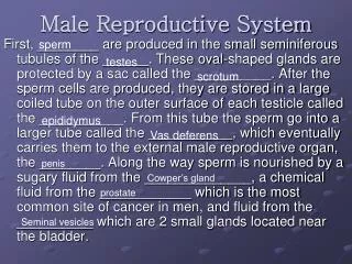
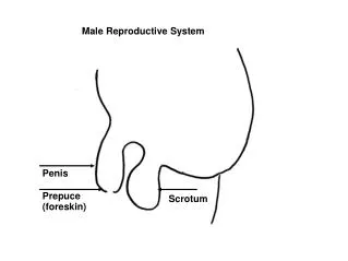
In male, reproductive system includes: Two testes, right and left. Two epididymis, right and left. Two deferent ducts, right and left. Two ejaculatory ducts, right and left. Two seminal vesicles, right and left. One prostate gland. One penis. Two bulbo-urethral glands, right and left.
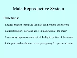
Testes are the primary sex organs. Here the male gametes are formed. Epididymis, deferent ducts and ejaculatory ducts transport mature male gametes (sperms) to the organ of copulation. Penis is the organ of copulation. Seminal vesicles, prostate gland and bulbo-urethral glands are associated accessory glandular structures.
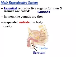
Testes • These are primary reproductive organs. • They are homologous to ovaries in female. • They are ellipsoidal shaped structures. • They are not in pelvis, different to ovaries. • They are included in external genitalia and suspended in the scrotum. • Left testis is usually about 1 cm lower than the right.
Each testis is compressed laterally, having upper and lower poles, medial and lateral surfaces and anterior and posterior borders. The anterior border is convex and posterior border is nearly straight. Average testicular dimensions are 4-5 cm in length, 2.5 cm breadth and 3 cm antero-posterior diameter. Their weight varies from 10.5-14 g.
COVERINGS OF TESTIS • Tunica Vaginalisis like peritoneum. It covers the whole testis except posterior border. • Tunica albugineais a dense irregular connective tissue. It forms the true capsule of testis. • Tunica Vasculosais present internal to tunica albuginea.
Microscopic picture of testis showing three coats of testis.
MICROSCOPIC STRUCTURE • There are 200-300 lobules in each testis. • Each lobule contains 1-3 horseshoe shaped highly convoluted seminiferoustubules embedded in loose connective tissue. • This loose connective tissue contains groups of interstitial cells ofLeydig outside the seminiferous tubules.
SEMINIFEROUS TUBULES • Highly convoluted tubules. About 0.2mm in diameter and 50cm in length. Horseshoe shaped. The ends are straight. Here they are given the name straight tubules. The straight tubules open into the rete testis. • Each seminiferous tubule is lined by complex stratified epithelium called seminiferous epithelium. The epithelium rests on a thin basal lamina, which is covered by connective tissue containing fibroblasts, numerous fibers and myoid cells.
Sustentacular Cells of Sertoli. Spermatogenic Cells
Sustentacular Cells of Sertoli • These are tall columnar cells resting on basal lamina of seminiferous tubules. The cell outline is very irregular and indistinct. They have invaginations on their basal surfaces, their sidewalls and their luminal surfaces. • The nucleus is towards the base. It is located some distance above the base of the cell. It is pale and ovoid. It contains a nucleolus.
The adjacent surfaces show complex occluding junctions. • These junctions are near the bases and constitute blood-testis barrier.
Spermatogenic Cells • These are germ cells. • They form a stratified layer of epithelium consisting of four to eight cell types.
Spermatogonia: • Until puberty these are the only germ cell type present. They occupy a space present between the basal lamina and sertoli cells. They contain 46 chromosomes. • Three basic types of spermatogonia are: • Type A dark spermatogonia • Type A pale spermatogonia • Type B spermatogonia • Spermatogonia multiply by mitosis.
Primary Spermatocytes: Some of the spermatogonia cells enlarge and differentiate into primary spermatocytes. They are the largest germ cells seen within seminiferous tubules. Initially they are in the basal compartment of seminiferous tubule. Then they cross blood-testis barrier and lie between the adjacent Sertoli cells. Here they are in the invaginations present in the lateral walls of Sertoli cells Each cell is spherical or ovoid in outline. They contain 46 chromosomes.
Secondary Spermatocytes: • Primary spermatocytes undergo first meiotic division and form secondary spermatocytes. • These are about half the size of primary spermatocytes. • They lie in the invaginations in the lateral walls of Sertoli cells near the lumen of the seminiferous tubules. • They contain 23 chromosomes.
Spermatids: • Secondary spermatocytes undergo second meiotic division and form spermatids. These are about the size of secondary spermatocytes and contain 23 chromosomes. • They migrate to the luminal surface of Sertoli cell and lie in the invaginations there. • Here they transform into spermatozoa by a process called spermiogenesis.
Spermatozoa: • Each spermatozoon is 60 µm in length • It consists of head, neck, middle piece and tail. • Head is 4µm in length, 3 µm in width and 1 µm in thickness • It has • a nucleus • with a cap called acrosome • surrounded by cell membrane
Middle pieceis separated from head by a narrow neck. It is 7 µm in length and 1 µm in diameter It contains Axoneme a core of longitudinal filaments 2+9 arrangement surrounded by nine dense fibers surrounded by a sheath of mitochondria Surrounded by cell membrane
Tail has two parts Principle piece is 40 µm in length. It consists of Axoneme. It has two central and nine peripheral double filaments Seven outer dense fibers Fibrous sheath Cell membrane End piece (8 µm in length)has axoneme and cell membrane only
Heads are embedded in the invaginations on luminal surfaces of Sertoli cells • and tails float in the lumen of seminiferous tubules.
Sperms: • When the spermatozoa become detached from Sertoli cell and free in the lumen of seminiferous tubules they are called sperms.
The interstitial tissue, within the lobules is loose connective tissue containing. • Cells. Fibroblasts, macrophages, mast cells, undifferentiated mesenchymal cells and interstitial cells of Leydig. • Collagen fibers. • Blood and lymph vessels. • Nerves.
The specific interstitial cells of Leydig are a marked feature of this tissue. They lie in groups. They are large rounded cells contain central rounded nucleus. The nucleus contains course chromatin granules and a distinct nucleolus. Under electron microscopy my dear students you will observe abundant smooth endoplasmic reticulum. Interstitial cells of Leydig secrete testosterone.
Smooth endoplasmic reticulum is concerned with synthesis of steroid hormone. • Rough endoplasmic reticulum is concerned with synthesis of proteins.
Spermatogenic Cells • Spermatogonia: 46Mitosis • Primary Spermatocytes: 46Meiosis I • Secondary Spermatocytes: 23Meiosis II • Spermatids: 23Spermeogenesis • Spermatozoa: 23Attached • Sperms: 23Free
Spermatogenic cells • Begin sperm production at puberty • Sertoli or sustentacular cells • Junctions form blood-testis barrier • Nourish spermatogenic cells • Carry out phagocytosis • Control spermatogenic movement • Produce fluid for transport • Secrete hormone inhibin • Leydig (interstitial) cells • Secrete testosterone