Download

1 / 15
150 likes | 414 Views
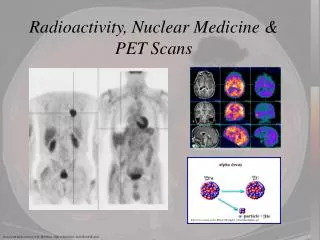
Brain PET Scans in Patients with Minimal Pain . Asokumar Buvanendran, MD, Amjad Ali, MD, Travis R. Stoub, PhD, Jeffrey S. Kroin, Ph.D., Kenneth J. Tuman, M.D. . Departments of Anesthesiology, Radiology, and Neurology Rush University Medical Center Rush Medical College Chicago, IL .

E N D
Brain PET Scans in Patients with Minimal Pain Asokumar Buvanendran, MD, Amjad Ali, MD, Travis R. Stoub, PhD, Jeffrey S. Kroin, Ph.D., Kenneth J. Tuman, M.D. Departments of Anesthesiology, Radiology, and Neurology Rush University Medical Center Rush Medical College Chicago, IL
Introduction • Positron emission tomography (PET) is an imaging technique that can quantify increases in nerve cell activity in selective regions of the brain. • Earlier studies have examined the pattern of increased brain activity that follows experimentally-induced acute pain.1,2,3 1Brain 1998;121:931. 2J Neurophysiol 1999; 82:1934. 3Anesth Analg 2007; 105:1784.
Is Seeing Believing? Can Pain Be Measured?
Increased Activity in the Brain with Pain • 6 different areas are being studied: • Anterior Cingulate Cortex (ACC) • Primary Somatosenory Cortex (S1) • Secondary Somatosensory Cortex (S2) • Insular Cortex (IC) • Pre-frontal Cortex (PF) • Thalamus
Moderate Postoperative Pain and Brain Activation After TKA • Baseline MRI of the brain and PET scan • Epidural Anesthesia and Analgesia for postoperative pain • Stopped epidural: Moderate pain → PET • Epidural infusing: No Pain → PET
Postoperative Moderate Pain after Left TKA Contralateral Somatosensory Cortex Right side Precuneus Thalamus
Current Study • However, baseline PET activity (pre-stimulation or pre-surgery) in the brain has not been examined in detail. • Knowledge of baseline activity in patients with no pain or mild pain prior to surgical or anesthetic intervention is important for future research involving PET assessment of modulation of postoperative pain. • In this study we examine PET brain activation at rest in six patients with minimal pain.
Methods • In a quiet room with low ambient light, fasted patients were injected intravenously with the PET radionuclide 18F-fluoro-2-deoxyglucose. • After waiting at least 30 min, a brain PET scan was performed. • Prior to each PET scan, pain scores were measured using the verbal rating scale, with 0=no pain, 10=worst imaginable pain.
Analysis • For the patients, digital files of PET scans were co-localized with a standard stereotaxic MRI. • To compare PET scans among different patients, a linear normalization was applied by dividing regional activity by whole brain activity for each scan.1 • A relevant increase in glucose metabolism was assumed if more than 50 adjacent voxels showed a Z score > 2.2 1Eur Neuropsychopharm 2002;12:527-44. 2 JNNP 2003; 74:922
Results The mean baseline pain score in the six patients was 1.17 (range 0-2). PET activation in these subjects was primarily in the occipital lobe, and putamen bilaterally Putamen Occipital lobe
There was no significant activation in pain-related regions Somatosensory cortex (SI or SII) SI SII
There was no significant activation in pain-related regions Anterior cingulate gyrus (ACC) ACC
There was no significant activation in pain-related regions Insula Also, no activation in thalamus
Discussion • To fully comprehend acute, chronic, and postoperative pain PET studies, baseline brain activation must be characterized. • In patients reporting minimal pain, we have established that there was no brain activation in pain-related regions. • This suggests that changes in brain activation after intervention (e.g. joint replacement surgery) will not be obscured by the patient’s baseline brain activity as long as the baseline pain score is 2 or less.
Some activity in superior temporal gyrus and frontal cortex: Not Pain Activated site Frontal cortex Sup Temporal Gyrus